. 2013 Oct-Dec;4(4):208-20.
doi: 10.4161/sgtp.26602. Epub 2013 Oct 22.

Multiple cytoskeletal pathways and PI3K signaling mediate CDC-42-induced neuronal protrusion in C. elegans

Affiliations

Multiple cytoskeletal pathways and PI3K signaling mediate CDC-42-induced neuronal protrusion in C. elegans

Jamie K Alan et al. Small GTPases. 2013 Oct-Dec.

Abstract

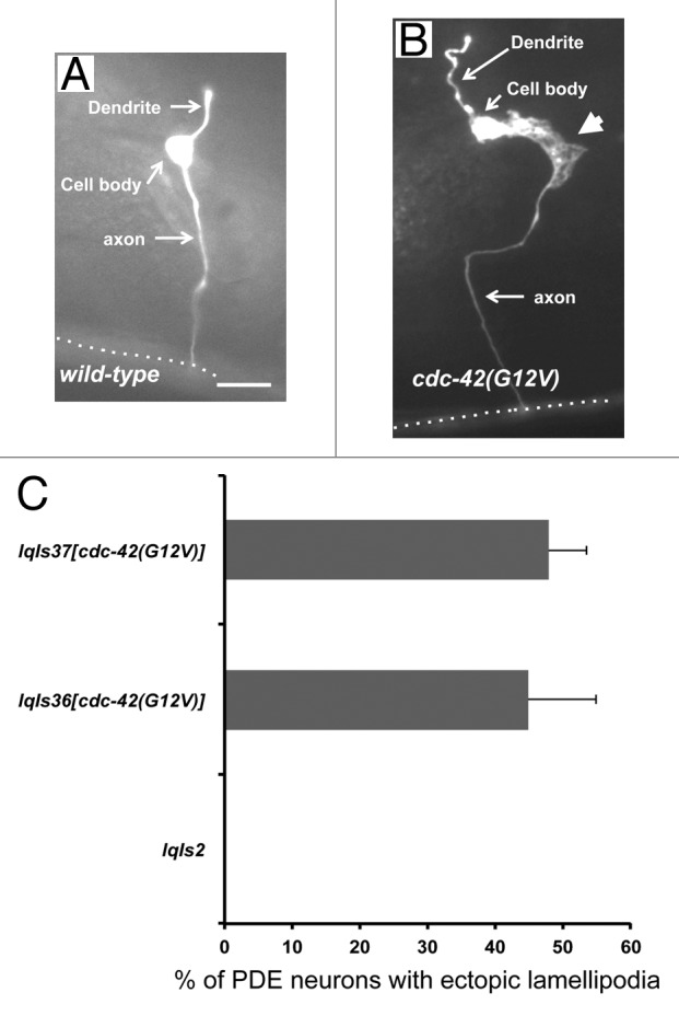
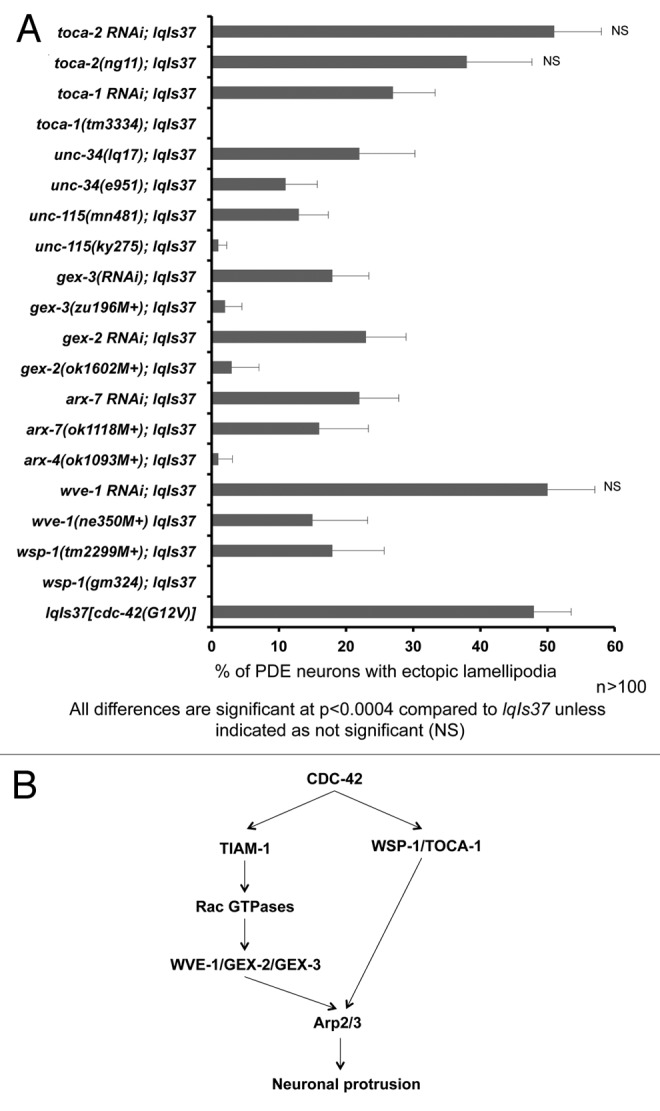
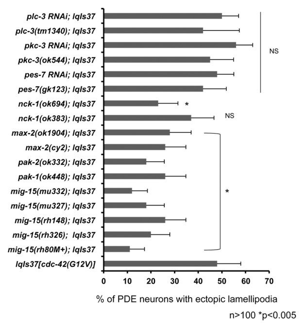
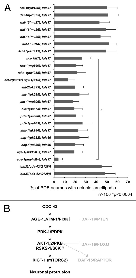
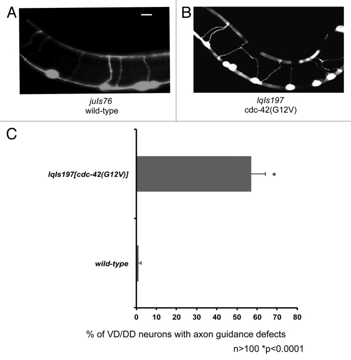
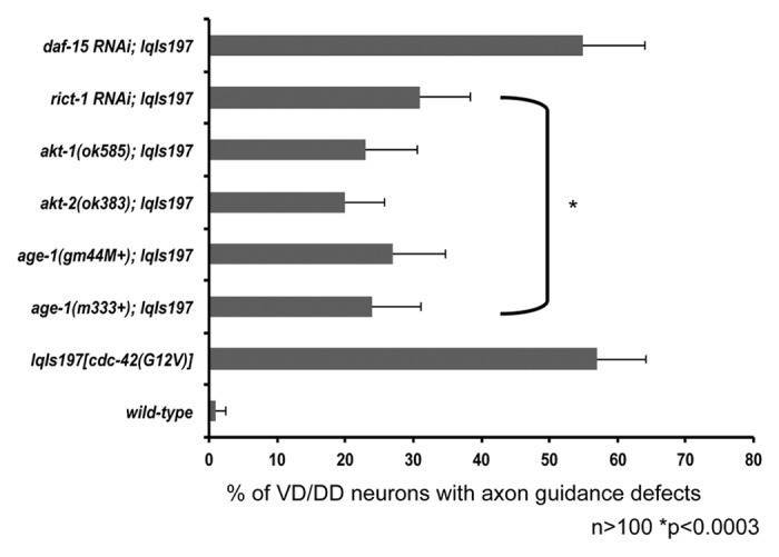
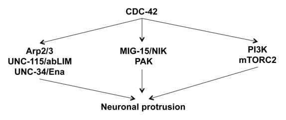
Rho GTPases are key regulators of cellular protrusion and are involved in many developmental events including axon guidance during nervous system development. Rho GTPase pathways display functional redundancy in developmental events, including axon guidance. Therefore, their roles can often be masked when using simple loss-of-function genetic approaches. As a complement to loss-of-function genetics, we constructed a constitutively activated CDC-42(G12V) expressed in C. elegans neurons. CDC-42(G12V) drove the formation of ectopic lamellipodial and filopodial protrusions in the PDE neurons, which resembled protrusions normally found on migrating growth cones of axons. We then used a candidate gene approach to identify molecules that mediate CDC-42(G12V)-induced ectopic protrusions by determining if loss of function of the genes could suppress CDC-42(G12V). Using this approach, we identified 3 cytoskeletal pathways previously implicated in axon guidance, the Arp2/3 complex, UNC-115/abLIM, and UNC-43/Ena. We also identified the Nck-interacting kinase MIG-15/NIK and p21-activated kinases (PAKs), also implicated in axon guidance. Finally, PI3K signaling was required, specifically the Rictor/mTORC2 branch but not the mTORC1 branch that has been implicated in other aspects of PI3K signaling including stress and aging. Our results indicate that multiple pathways can mediate CDC-42-induced neuronal protrusions that might be relevant to growth cone protrusions during axon pathfinding. Each of these pathways involves Rac GTPases, which might serve to integrate the pathways and coordinate the multiple CDC-42 pathways. These pathways might be relevant to developmental events such as axon pathfinding as well as disease states such as metastatic melanoma.

Keywords: AGE-1/PI3K; Arp2/3; CDC-42; MIG-15; UNC-115; axon pathfinding; mTORC2.


Figures

None
Figure 1. Expression of CDC-42(G12V) results in the formation of ectopic lamellipodia in PDE neurons. (A) A micrograph of a PDE neuron of a WT adult animal (B) A PDE neuron of an adult animal expressing CDC-42(G12V). An ectopic lamellipodial protrusion is indicated by the large arrowhead. The dotted line indicates the ventral nerve cord. The scale bar represents 5 μm. (C) Quantitation of PDE defects. lqIs2 is the osm-6::gfp control transgene and lqIs36 and lqIs37 are the Posm-6::CDC-42(G12V) activated CDC-42 transgenes. At least 100 neurons were scored for each genotype. p < 0.0001 as determined by Fisher Exact Analysis. The error bars represent 2x the standard error of the proportion (2x SEP).
None
Figure 2. Multiple cytoskeletal pathways suppress activated CDC-42 in PDE neurons. (A) Quantitation of PDE defects. lqIs37 is Posm-6::CDC-42(G12V) activated CDC-42 transgene. The Y-axis denotes the genotype and the X-axis represents the percentage of ectopic lamellipodia formation. “M+” denotes that the animals had wild-type maternal contribution. The number of axons scored > 100 for each genotype. p < 0.0004 as determined by Fisher Exact Analysis. The error bars represent 2x SEP. (B) A diagram depicting CDC-42 signaling to the Arp2/3 complex based on genetic results in this study and in other work.
None
Figure 3. MIG-15(NIK) and p21 activated kinase (PAK) suppress activated CDC-42 in PDE neurons. Quantitation of PDE defects. lqIs2 is the osm-6::gfp control transgene and lqIs37 is Posm-6::CDC-42(G12V) activated CDC-42 transgene. The Y-axis denotes the genotype and the X-axis represents the percentage of ectopic lamellipodia formation. “M+” denotes that the animals had wild-type maternal contribution. The number of axons scored > 100. *p < 0.005 as determined by Fisher Exact Analysis. The error bars represent 2x SEP.
None
Figure 4. CDC-42 utilizes specific components of the PI3K pathway to drive ectopic lamellipodia formation in PDE neurons. (A) PI3K signaling mutants suppress CDC-42(G12V). The Y-axis denotes the genotype and the X-axis represents the percentage of ectopic lamellipodia formation. “M+” denotes that the animals had wild-type maternal contribution. The number of axons scored > 100. *p < 0.0004 as determined by Fisher Exact Analysis. The error bars represent 2x SEP. (B) A diagram of the genetic interaction with CDC-42 and the PI3K signaling pathway. Molecules in gray are those not involved in CDC-42-induced neuronal protrusion.
None
Figure 5. An activated allele of akt-1 results in the formation of ectopic lamellipodia, similar to activated CDC-42 (A) A micrograph of a PDE neuron of a WT adult animal. (B) A PDE neuron of an akt-1(gm144) adult animal. An ectopic lamellipodial protrusion is indicated by the arrow. The scale bars represents 5 μm. (C) Quantitation of PDE defects. lqIs2 is the osm-6::gfp control transgene. The Y-axis denotes the genotype and the X-axis represents the percentage of ectopic lamellipodia formation. The number of axons scored > 100. *p < 0.0001 as determined by Fisher Exact Analysis. The error bars represent 2x SEP.
None
Figure 6. An activated allele of akt-1 is suppressed by components of the mTORC2 complex, but not the mTORC1 complex. Quantitation of PDE defects. lqIs2 is the Posm-6::gfp control transgene. rict-1 is in the mTORC2 complex and daf-15 is in the mTORC1 complex. The Y-axis denotes the genotype and the X-axis represents the percentage of ectopic lamellipodia formation. The number of axons scored > 100. *p < 0.0004 as determined by Fisher Exact Analysis. The error bars represent 2x SEP.
None
Figure 7. AGE-1 and RICT-1 act cell autonomously in the PDE neuron. Transgenic lines containing cell-specific RNAi against, age-1, rict-1 and daf-15 were constructed and crossed with lqIs37. All of the strains are in a sid-1(pk3321) background, which limits RNAi spreading. The Y-axis denotes the genotype and the X-axis represents the percentage of ectopic lamellipodia formation. The number of axons scored > 100. *p < 0.005 as determined by Fisher Exact Analysis. The error bars represent 2x SEP.
None
Figure 8. Expression of CDC-42(G12V) results in the axon guidance defects in the VD/DD motor neurons. (A) A micrograph of transgenic animals expressing Punc-25::gfp. (B) A transgenic animal expressing Punc-25::CDC-42(G12V) with axon guidance defects. The scale bars represent 5 μm. (B) Quantitation of VD/DD defects. juIs76 is the Punc-25::gfp control transgene. The Y-axis denotes the genotype and the X-axis represents the percentage of axon pathfinding defects. The number of axons scored > 100. *p < 0.0001 determined by Fisher Exact Analysis. The error bars represent 2x SEP.
None
Figure 9. Components of the AGE-1 pathway and the mTORC2 complex suppress the axon guidance defects driven by activated CDC-42 in the VD/DD motor neurons. Quantitation of axon guidance defects in the VD/DD motor neurons. Null alleles several downstream CDC-42 effectors including: age-1, akt-1, and akt-2 suppress the axon pathfinding defects driven by activated CDC-42 in VD/DD motor neurons. Components of the mTORC2 pathway (rict-1) but not the mTORC2 pathway (daf-15) suppress the axon pathfinding defects driven by CDC-42(G12V). The Y-axis denotes genotype and the X-axis represents percentage of axon pathfinding defects. The number of axons scored > 100. *p < 0.0003 determined by Fisher Exact Analysis. The error bars represent 2x SEP.
None
Figure 10. Multiple pathways mediate CDC-42-induced neuronal protrusion. A diagram of the multiple pathways mediating CDC-42(G12V)-induced neuronal protrusion described in this work.

References

    1. Lundquist EA. Rac proteins and the control of axon development. Curr Opin Neurobiol. 2003;13:384–90. doi: 10.1016/S0959-4388(03)00071-0. - DOI - PubMed
    1. Luo L. Rho GTPases in neuronal morphogenesis. Nat Rev Neurosci. 2000;1:173–80. doi: 10.1038/35044547. - DOI - PubMed
    1. Dickson BJ. Rho GTPases in growth cone guidance. Curr Opin Neurobiol. 2001;11:103–10. doi: 10.1016/S0959-4388(00)00180-X. - DOI - PubMed
    1. Sulston J. J H. The nematode Caenorhabditis elegans. Cold Spring Harbor, NY: Cold Spring Harbor Laboratory Press, 1988. - PubMed
    1. Etienne-Manneville S, Hall A. Rho GTPases in cell biology. Nature. 2002;420:629–35. doi: 10.1038/nature01148. - DOI - PubMed

Publication types

MeSH terms

Substances