

Sprouting and intussusceptive angiogenesis in postpneumonectomy lung growth: mechanisms of alveolar neovascularization
- PMID: 24150281
- PMCID: PMC4061467
- DOI: 10.1007/s10456-013-9399-9
Sprouting and intussusceptive angiogenesis in postpneumonectomy lung growth: mechanisms of alveolar neovascularization
Abstract
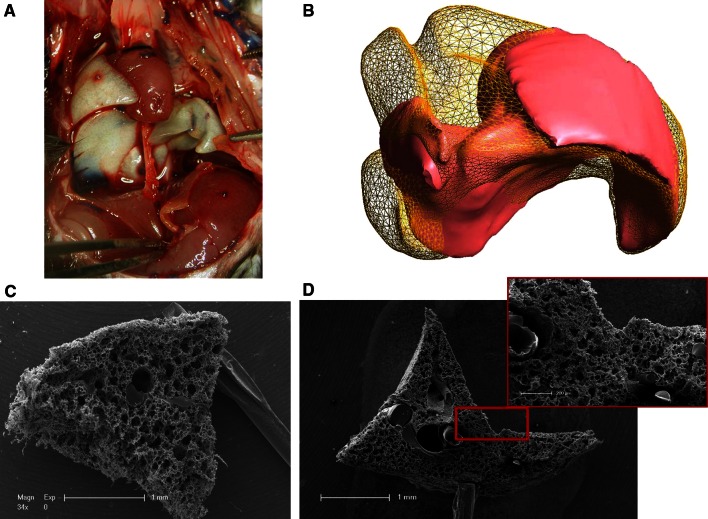
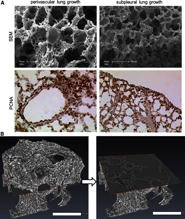
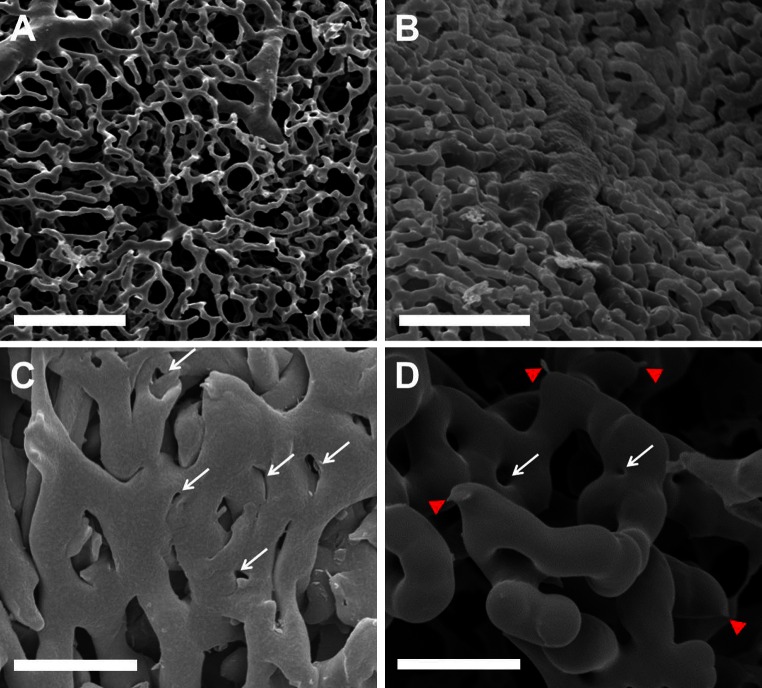
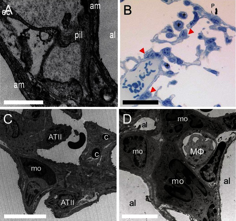
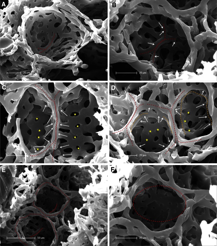
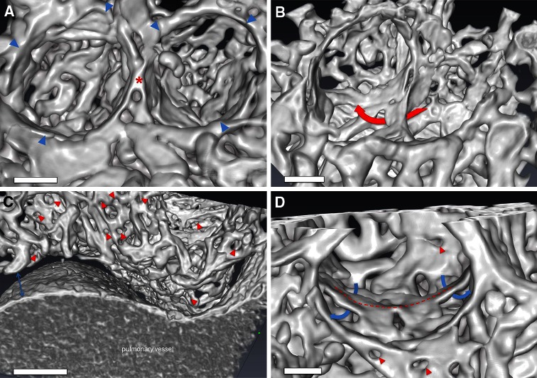
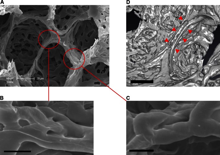
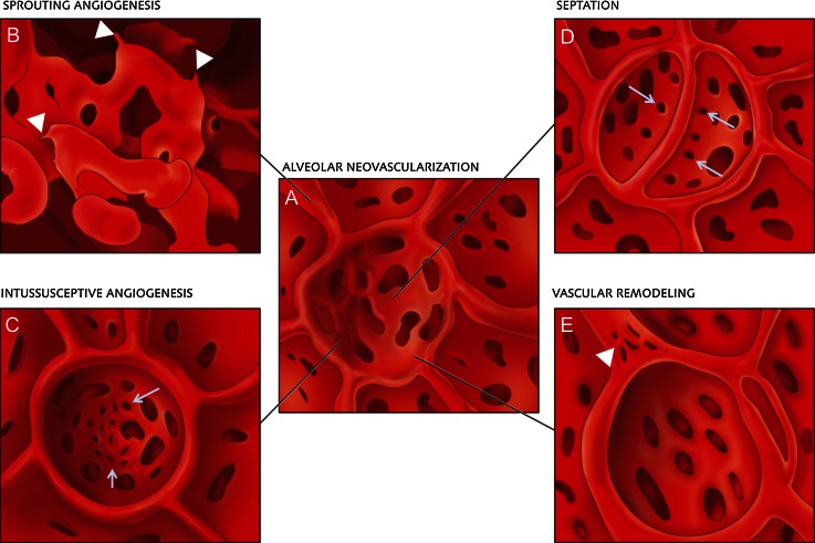
In most rodents and some other mammals, the removal of one lung results in compensatory growth associated with dramatic angiogenesis and complete restoration of lung capacity. One pivotal mechanism in neoalveolarization is neovascularization, because without angiogenesis new alveoli can not be formed. The aim of this study is to image and analyze three-dimensionally the different patterns of neovascularization seen following pneumonectomy in mice on a sub-micron-scale. C57/BL6 mice underwent a left-sided pneumonectomy. Lungs were harvested at various timepoints after pneumonectomy. Volume analysis by microCT revealed a striking increase of 143 percent in the cardiac lobe 14 days after pneumonectomy. Analysis of microvascular corrosion casting demonstrated spatially heterogenous vascular densitities which were in line with the perivascular and subpleural compensatory growth pattern observed in anti-PCNA-stained lung sections. Within these regions an expansion of the vascular plexus with increased pillar formations and sprouting angiogenesis, originating both from pre-existing bronchial and pulmonary vessels was observed. Also, type II pneumocytes and alveolar macrophages were seen to participate actively in alveolar neo-angiogenesis after pneumonectomy. 3D-visualizations obtained by high-resolution synchrotron radiation X-ray tomographic microscopy showed the appearance of double-layered vessels and bud-like alveolar baskets as have already been described in normal lung development. Scanning electron microscopy data of microvascular architecture also revealed a replication of perialveolar vessel networks through septum formation as already seen in developmental alveolarization. In addition, the appearance of pillar formations and duplications on alveolar entrance ring vessels in mature alveoli are indicative of vascular remodeling. These findings indicate that sprouting and intussusceptive angiogenesis are pivotal mechanisms in adult lung alveolarization after pneumonectomy. Various forms of developmental neoalveolarization may also be considered to contribute in compensatory lung regeneration.
Figures
Similar articles
-
Spatial dependence of alveolar angiogenesis in post-pneumonectomy lung growth.Angiogenesis. 2012 Mar;15(1):23-32. doi: 10.1007/s10456-011-9236-y. Epub 2011 Oct 4. Angiogenesis. 2012. PMID: 21969134 Free PMC article.
-
Remodeling of alveolar septa after murine pneumonectomy.Am J Physiol Lung Cell Mol Physiol. 2015 Jun 15;308(12):L1237-44. doi: 10.1152/ajplung.00042.2015. Epub 2015 Apr 10. Am J Physiol Lung Cell Mol Physiol. 2015. PMID: 26078396 Free PMC article.
-
Alveolar epithelial dynamics in postpneumonectomy lung growth.Anat Rec (Hoboken). 2013 Mar;296(3):495-503. doi: 10.1002/ar.22659. Anat Rec (Hoboken). 2013. PMID: 23408540 Free PMC article.
-
Intussusceptive angiogenesis: expansion and remodeling of microvascular networks.Angiogenesis. 2014 Jul;17(3):499-509. doi: 10.1007/s10456-014-9428-3. Epub 2014 Mar 26. Angiogenesis. 2014. PMID: 24668225 Free PMC article. Review.
-
Intussusceptive angiogenesis: its emergence, its characteristics, and its significance.Dev Dyn. 2004 Nov;231(3):474-88. doi: 10.1002/dvdy.20184. Dev Dyn. 2004. PMID: 15376313 Review.
Cited by
-
Microvascular Skeletal-Muscle Crosstalk in Health and Disease.Int J Mol Sci. 2023 Jun 21;24(13):10425. doi: 10.3390/ijms241310425. Int J Mol Sci. 2023. PMID: 37445602 Free PMC article. Review.
-
The puzzling mechanism of compensatory lung growth.Stem Cell Investig. 2018 Mar 31;5:8. doi: 10.21037/sci.2018.03.01. eCollection 2018. Stem Cell Investig. 2018. PMID: 29682515 Free PMC article. No abstract available.
-
A method for evaluating the murine pulmonary vasculature using micro-computed tomography.J Surg Res. 2017 Jan;207:115-122. doi: 10.1016/j.jss.2016.08.074. Epub 2016 Aug 31. J Surg Res. 2017. PMID: 27979466 Free PMC article.
-
Evidence for pleural epithelial-mesenchymal transition in murine compensatory lung growth.PLoS One. 2017 May 19;12(5):e0177921. doi: 10.1371/journal.pone.0177921. eCollection 2017. PLoS One. 2017. PMID: 28542402 Free PMC article.
-
The influence of geranylgeraniol on microvessel sprouting after bisphosphonate substitution in an in vitro 3D-angiogenesis assay.Clin Oral Investig. 2017 Apr;21(3):771-778. doi: 10.1007/s00784-016-1842-z. Epub 2016 May 12. Clin Oral Investig. 2017. PMID: 27170294
References
-
- U.S. Department of Health and Human Services. National Institutes of Health. National Heart Lung and Blood Institute. Morbidity and Mortality (2012) Chartbook on Cardiovascular, Lung and Blood Diseases
Publication types
MeSH terms
Grants and funding
LinkOut - more resources
Full Text Sources
Other Literature Sources
Miscellaneous
