. 2013 Oct 22:9:699.
doi: 10.1038/msb.2013.43.

Sequential induction of auxin efflux and influx carriers regulates lateral root emergence

Affiliations

Sequential induction of auxin efflux and influx carriers regulates lateral root emergence

Benjamin Péret et al. Mol Syst Biol. .

Abstract

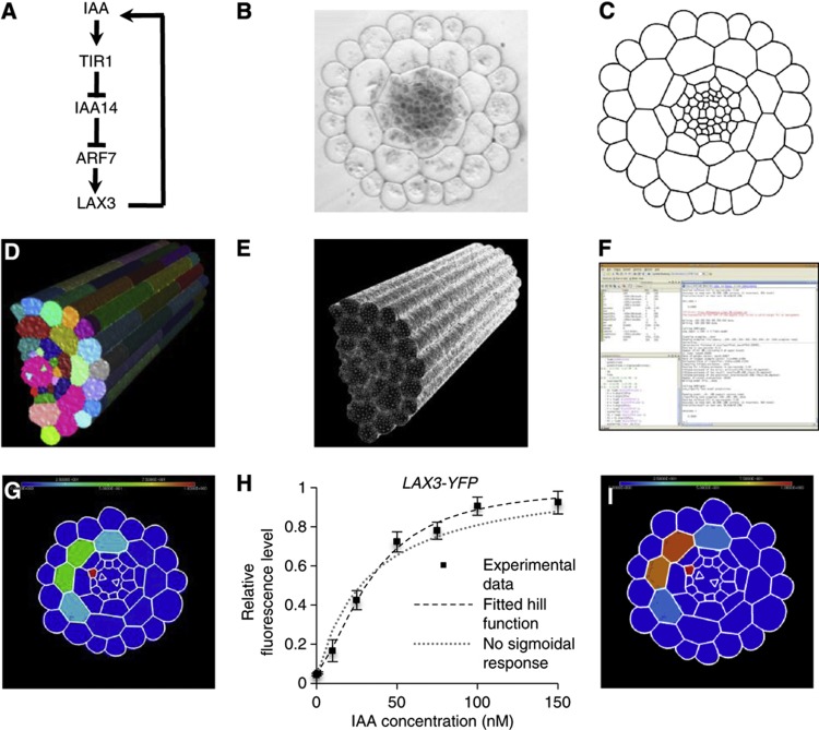
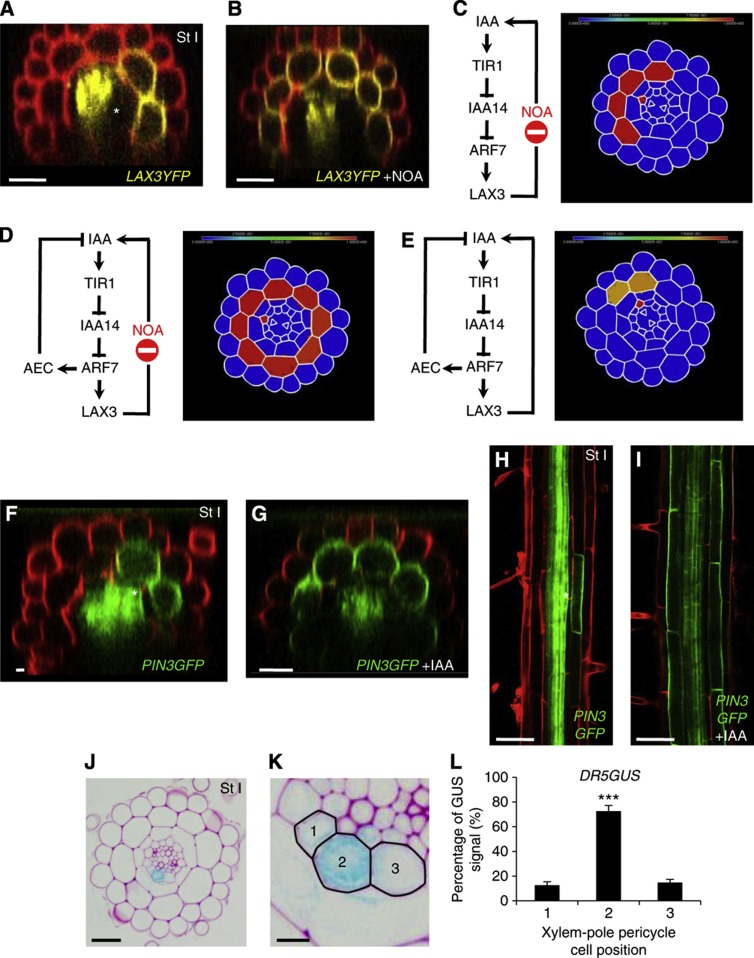
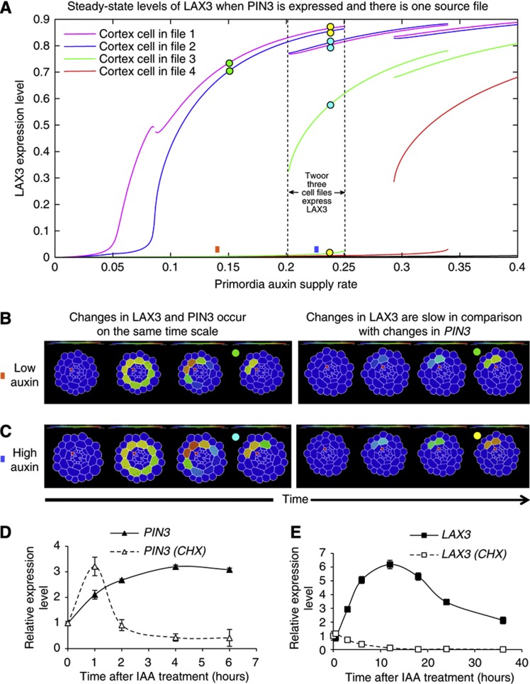
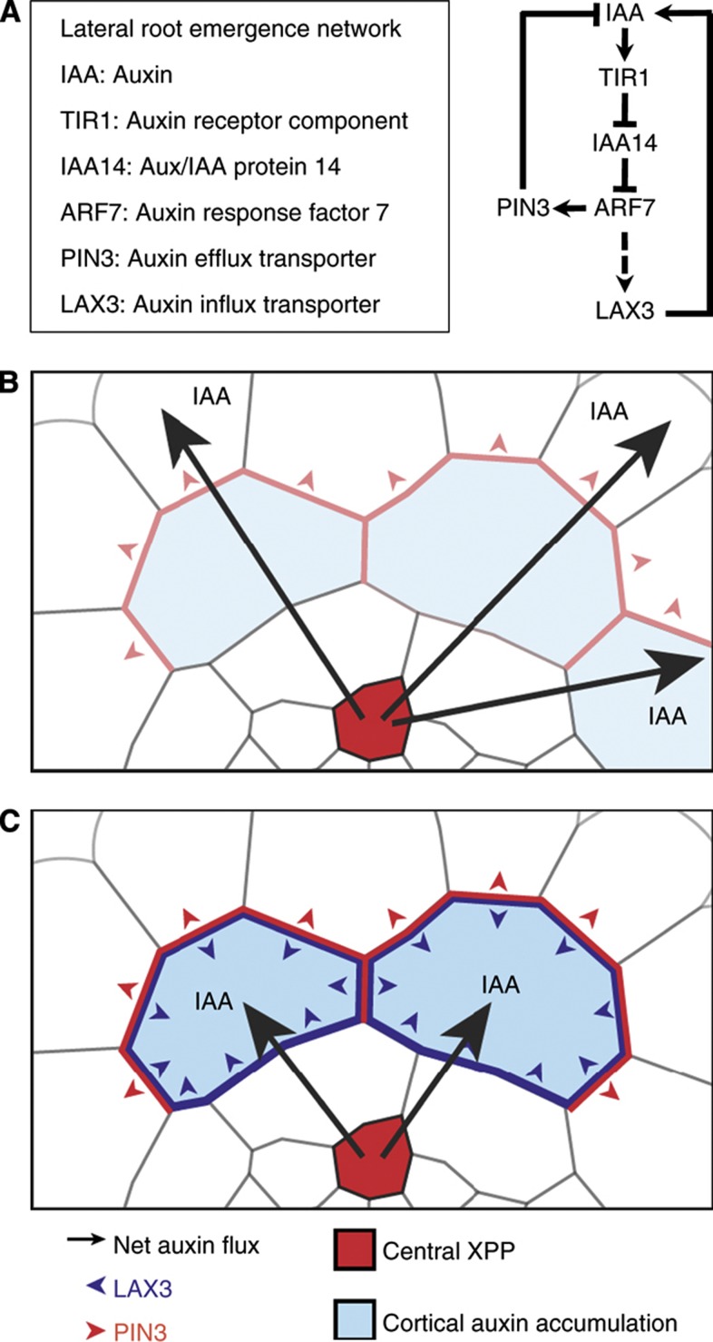
In Arabidopsis, lateral roots originate from pericycle cells deep within the primary root. New lateral root primordia (LRP) have to emerge through several overlaying tissues. Here, we report that auxin produced in new LRP is transported towards the outer tissues where it triggers cell separation by inducing both the auxin influx carrier LAX3 and cell-wall enzymes. LAX3 is expressed in just two cell files overlaying new LRP. To understand how this striking pattern of LAX3 expression is regulated, we developed a mathematical model that captures the network regulating its expression and auxin transport within realistic three-dimensional cell and tissue geometries. Our model revealed that, for the LAX3 spatial expression to be robust to natural variations in root tissue geometry, an efflux carrier is required--later identified to be PIN3. To prevent LAX3 from being transiently expressed in multiple cell files, PIN3 and LAX3 must be induced consecutively, which we later demonstrated to be the case. Our study exemplifies how mathematical models can be used to direct experiments to elucidate complex developmental processes.


Conflict of interest statement

The authors declare that they have no conflict of interest.

Figures

Figure 1
Figure 1
Lateral root (LR) formation and emergence in Arabidopsis thaliana. (A) Cross-section of an Arabidopsis root (during stages 0–I of LRP emergence) showing the different cell types, with the position of the cross-section shown in (B). Xylem-pole pericycle cells are grouped in three cell files and are in contact with several endodermal cell files, which in turn about several cortical cell files (highlighted cells). (B) Stages of LR formation. Between stages 0 and I, the XPP cells (from which the LRP originate) undergo several rounds of anticlinal division. Note that in the transverse direction cells vary in length and appear in a staggered formation. (C, D) LAX3 protein accumulation pattern was visualised using a functional pLAX3:LAX3YFP fusion in a tangential root section (C) or a cross-section (D) (with the position of the cross-section shown indicated by the dashed line in (C)). The asterisk highlights the position of one of the XPP cells, from which the LRP originate. (EG) Statistical analysis was performed on lateral root primordia of plants bearing a functional pLAX3:LAX3YFP fusion (n=40) to determine the number of files (E), the number of cortical cells per file (F) and the total number of cortical cells (G) showing LAX3 expression.
Figure 2
Figure 2
The primordium acts as a source of auxin to induce genes involved in LR emergence. (AG) The PIN2 auxin efflux carrier removes auxin from the apex of the LR primordium (A). As a result of the overaccumulation of auxin in the apex of the LRP in the pin2 mutant background, both LAX3 (B) and PG (C) are induced. Overaccumulation of auxin in the LRP tip in the pin2 mutant background increases the strength but does not alter the expression pattern of pLAX3:GUS (D, E) and pPG:GUS (F, G). Asterisks indicate a significant difference with corresponding control experiment by Student’s t-test (*P<0.05; **P<0.01). Bars are 50 μm (DG).
Figure 3
Figure 3
Auxin moves from the pericycle cells towards the outer tissues. (AL) Tissue-specific expression of the Agrobacterium indole-3-acetamide hydrolase (iaaH) gene was used as a tool to control auxin synthesis. Expression of the iaaH gene under the control of the J0121 GAL4 driver line (B) resulted in production of numerous lateral roots upon 1 μM indole-3-acetamide treatment whereas wild-type plants (Col-0) were not affected (A). Transgenic Arabidopsis plants were engineered to synthesise IAA in xylem-pole pericycle (XPP) cells using the XPP-specific GAL4 drive line, J0121 (C) by targeting the expression of the bacterial iaaH enzyme fused to the RFP tag (D). Therefore, the J0121 and iaaH fluorescent markers (respectively GFP and RFP) are co-expressed in the XPP tissue (E). Plant roots were fed with 5 μM deuterated indole-3-acetamide (D5-IAM), and the ratio of D5-IAA to IAA was determined by mass spectrometry analysis. A strong D5-IAA signal was detected upon 10 and 20 h after incubation in the J0121>>iaaH-RFP line, whereas very little D5-IAA was detected in the control (J0121) line even after 20 h of incubation (F). As a result of auxin synthesis in the xylem-pole pericycle cells, we observed an overexpression of LAX3 (G) and PG (H). The J0121 line is specifically expressed in three pericycle cell files associated with each of the xylem poles as seen with the GFP reporter (I) and as reported previously (Laplaze et al, 2005). (JL) We monitored the effect of auxin synthesis in the pericycle on the LAX3 expression pattern by crossing the J0121>>iaaH-RFP plants with the pLAX3:LAX3YFP-expressing plants and subsequent analysis on the F1 progeny. LAX3-YFP accumulation was visualised upon 18 h treatment with mock (J), 1 μM IAM (K) or 10μM IAM (L). Note that strong expression of LAX3 in the vasculature is seen as in the non-treated control. Bars are 25 μm (IL), 50 μm (CE) and 5 mm (A, B). Data shown are mean value±s.e.m. and n=100 plants. Asterisks indicate a significant difference with corresponding control experiment by Student’s t-test (*P<0.05; **P<0.01; ***P<0.001).
Figure 4
Figure 4
Computational 3D model of LAX3 induction by auxin in the cortical cells of the Arabidopsis root. (A) Representation of the gene regulatory network controlling lateral root emergence. IAA: auxin; TIR1: F-box protein that interacts with IAA14 to form the auxin receptor complex; IAA14: Aux/IAA protein acting as a negative regulator of ARF7; ARF7: Auxin response factor 7, a transcription factor that activates downstream auxin responsive genes; LAX3: auxin influx transporter. (BF) The meshing pipeline. A cross-section of a resin embedded wild-type (Col-0) Arabidopsis root (B) was manually digitised using Adobe Illustrator (C) and then extruded to realistically recreate the third dimension (including transverse walls, see main text) (D). A triangular mesh of the 3D object surface was generated using the CGAL library (E) and the surface triangles were imported into MATLAB to run the simulations (F). (G) Model output showing diffuse LAX3 protein accumulation in four cortical cell files (as indicated by a heatmap scale ranging from 0 (blue) to 1 (red); simulations shown are for the case in which the auxin source is provided by cells in a single XPP file; indicated in red). (H) LAX3 auxin dose response was determined by confocal microscopy of the LAX3-YFP protein and fitted to a Hill function (best fit: Hill Coefficient=2; threshold=40 nM); if no sigmoidal response was assumed (in the sense that the Hill Coefficient=1), then the fitted curve consistently overestimated the LAX3 response at low levels of auxin and underestimated it at high levels of auxin. Values are plotted as mean (±s.e., n=3). (I) Including the sigmoidal response increased the sharpness of the predicted LAX3 expression pattern, but still shows expression in four cortical cells. Scale bar is 25 μm (B).
Figure 5
Figure 5
Computational 3D modelling of LAX3 expression predicts the involvement of the auxin efflux transporter PIN3. (AD) Accumulation of LAX3-YFP in the non-treated conditions (A) or after 18 h treatment with 10 μM 2-NOA (B). Simulation of LAX3 induction in the presence of auxin influx inhibitor NOA in the absence of an auxin-induced efflux carrier (AEC) shows expression of LAX3 to be limited to a few cell files, in contrast to (B). In the presence of both an auxin influx inhibitor NOA and an AEC, LAX3 expression is predicted to spread throughout the cortex. When an AEC is included, the model can capture the stereotypical wild-type pattern (E). (FI) The auxin efflux carrier PIN3 is expressed in the cortical cells situated in front of the LRP and is induced by auxin in this tissue. Accumulation of PIN3GFP in the non-treated condition (F, H) and upon 18 h 1 μM IAA treatment (G, I). Asterisks indicate the presence of LRP in the non-treated conditions. IAA: auxin; TIR1: F-box protein that interacts with IAA14 to form the auxin receptor complex; IAA14: Aux/IAA protein acting as a negative regulator of ARF7; ARF7: Auxin response factor 7, a transcription factor that activates downstream auxin responsive genes; LAX3: auxin influx transporter. (JL) Cross-section of a DR5:GUS stained root showing a stage I primordium at low (J) and high magnification (K). Quantification of the DR5GUS signal (blue channel) in the three stained xylem-pole pericycle cells (numbered from 1 to 3 as shown in K) using ImageJ (L). Data shown are mean±s.e.m. and n=10 sections. Asterisks indicate a significant difference with corresponding control experiment by Student’s t-test (***P<0.001; n=10). Scale bars are 50 μm (H, I), 25 μm (F, G), 20 μm (J) and 5 μm (K).
Figure 6
Figure 6
PIN3 and LAX3 are sequentially induced to promote lateral root emergence. (A) Steady-state solutions to model version two (wherein both LAX3 and PIN3 are expressed, see Figure 5E and Table I) for various LRP auxin supply rates. For a supply rate within a certain range, LAX3 expression is restricted to just two cell files (as indicated). Within this range, the levels of LAX3 in other cell files remain low (compare with analogous plots obtained for model version one shown in Supplementary Modelling Figures M8–M10). For slightly higher supply rates, LAX3 can be expressed in three cell files at steady state. Overlap between the two regions corresponds to bistability. (B, C) Visualisation of model dynamics for various auxin supply rates (as indicated by square boxes) and LAX3 induction rates. Together, these determine the steady state to which the model tends (as indicated by the coloured circles). (B) If the auxin supply rate is chosen so that the model has one stable-steady state, and LAX3 and PIN3 are induced by auxin at the same rate, then LAX3 expression initially spreads circumferentially before being restricted to two cell files. (C) If the auxin supply rate is chosen so that the model is bistable, then LAX3 expression spreads throughout the cortex before being restricted to three cell files. In either case (bistable or monostable), if LAX3 is induced slowly by auxin, then the transient spread is eliminated and only two cell files are selected for LAX3 expression. (D, E) PIN3 but not LAX3 is a primary auxin response gene. Induction of PIN3 is observed from 1 h onward and is not blocked by treatment with the protein synthesis inhibitor cycloheximide (CHX) (D). Induction of LAX3 is detected from 4 h onward and is totally blocked by CHX treatment (E). Data shown are mean value±s.e.m. (n=100 plants).
Figure 7
Figure 7
Sequential action of PIN3 and LAX3 determines the specific expression pattern of LAX3 during lateral root emergence. (A) Regulatory network controlling LAX3 induction by auxin during lateral root emergence. (B) Auxin moves from the central xylem-pole pericycle file (XPP, red) towards the outer tissue and activates PIN3 in the cortex, resulting in a net flux of auxin towards the epidermis (red arrowheads). (C) The two files expressing PIN3 that make the most (indirect) contact with the cortex then accumulate enough auxin to induce LAX3 at a later stage (blue arrowheads). Accumulation of LAX3 then leads to further increases in auxin levels, which subsequently trigger cell separation and promote the passage of the LRP.

Comment in

  • Root systems analysis branches out.
    Palme K, Teale W. Palme K, et al. Mol Syst Biol. 2013 Oct 22;9:698. doi: 10.1038/msb.2013.51. Mol Syst Biol. 2013. PMID: 24150422 Free PMC article. No abstract available.

Similar articles

Cited by

References

    1. Andersen SU, Buechel S, Zhao Z, Ljung K, Novák O, Busch W, Schuster C, Lohmann JU (2008) Requirement of B2-type cyclin-dependent kinases for meristem integrity in Arabidopsis thaliana. Plant Cell 20: 88–100 - PMC - PubMed
    1. Benková E, Michniewicz M, Sauer M, Teichmann T, Seifertová D, Jürgens G, Friml J (2003) Local, efflux-dependent auxin gradients as a common module for plant organ formation. Cell 115: 591–602 - PubMed
    1. Blilou I, Xu J, Wildwater M, Willemsen V, Paponov I, Friml J, Heidstra R, Aida M, Palme K, Scheres B (2005) The PIN auxin efflux facilitator network controls growth and patterning in Arabidopsis roots. Nature 433: 39–44 - PubMed
    1. Ditengou FA, Teale WD, Kochersperger P, Flittner KA, Kneuper I, van der Graaff E, Nziengui H, Pinosa F, Li X, Nitschke R, Laux T, Palme K (2008) Mechanical induction of lateral root initiation in Arabidopsis thaliana. Proc Natl Acad Sci USA 105: 18818–18823 - PMC - PubMed
    1. Frey PJF (2001) Medit 3.0 interactive mesh visualization software http://www.ann.jussieu.fr/~frey/software.html

Publication types

MeSH terms