

Altered interhemispheric and temporal lobe white matter microstructural organization in severe chronic schizophrenia
- PMID: 24150571
- PMCID: PMC3924528
- DOI: 10.1038/npp.2013.294
Altered interhemispheric and temporal lobe white matter microstructural organization in severe chronic schizophrenia
Abstract
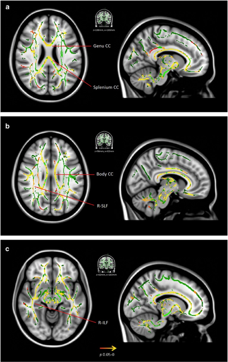
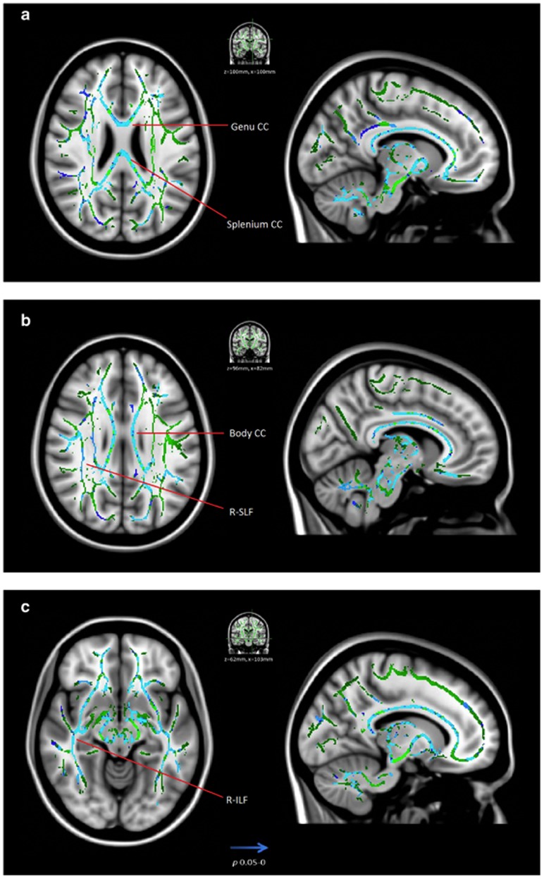
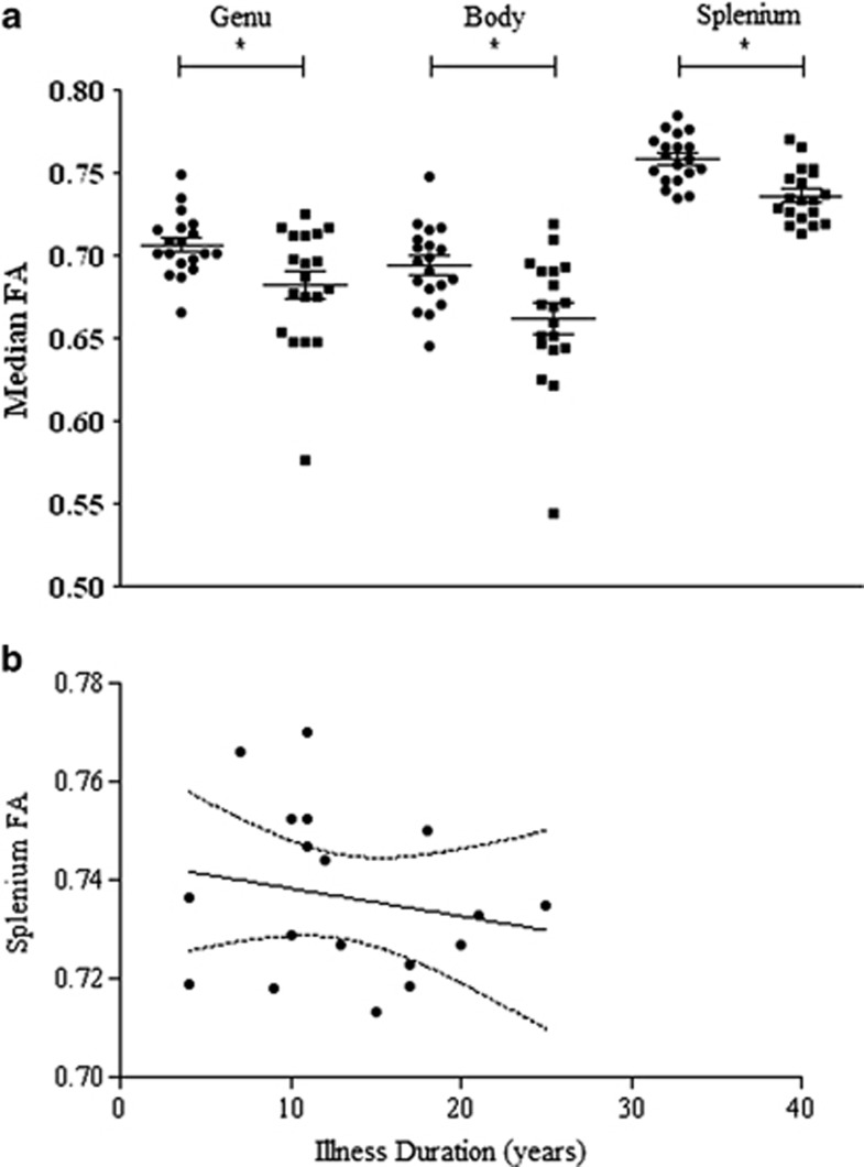
Diffusion MRI investigations in schizophrenia provide evidence of abnormal white matter (WM) microstructural organization as indicated by reduced fractional anisotropy (FA) primarily in interhemispheric, left frontal and temporal WM. Using tract-based spatial statistics (TBSS), we examined diffusion parameters in a sample of patients with severe chronic schizophrenia. Diffusion MRI data were acquired on 19 patients with chronic severe schizophrenia and 19 age- and gender-matched healthy controls using a 64 gradient direction sequence, (b=1300 s/mm(2)) collected on a Siemens 1.5T MRI scanner. Diagnosis of schizophrenia was determined by Diagnostic and Statistical Manual for Mental Disorders 4th Edition (DSM-IV) Structured Clinical Interview for DSM disorder (SCID). Patients were treatment resistance, having failed to respond to at least two antipsychotic medications, and had prolonged periods of moderate to severe positive or negative symptoms. Analysis of diffusion parameters was carried out using TBSS. Individuals with chronic severe schizophrenia had significantly reduced FA with corresponding increased radial diffusivity in the genu, body, and splenium of the corpus callosum, the right posterior limb of the internal capsule, right external capsule, and the right temporal inferior longitudinal fasciculus. There were no voxels of significantly increased FA in patients compared with controls. A decrease in splenium FA was shown to be related to a longer illness duration. We detected widespread abnormal diffusivity properties in the callosal and temporal lobe WM regions in individuals with severe chronic schizophrenia who have not previously been exposed to clozapine. These deficits can be driven by a number of factors that are indistinguishable using in vivo diffusion-weighted imaging, but may be related to reduced axonal number or packing density, abnormal glial cell arrangement or function, and reduced myelin.
Figures
References
-
- Agartz I, Andersson JL, Skare S. Abnormal brain white matter in schizophrenia: a diffusion tensor imaging study. Neuroreport. 2001;12:2251–2254. - PubMed
-
- Alexander AL, Hasan KM, Lazar M, Tsuruda JS, Parker DL. Analysis of partial volume effects in diffusion-tensor MRI. Magn Reson Med. 2001;45:770–780. - PubMed
-
- Anderson JLR, Jenkinson M, Smith S.2007. Non-linear registration, aka Spatial normalisation FMRIB technical report TR07JA2 www.fmrib.ox.ac.uk/analysis/techrep FMRIB Centre, Oxford, United Kingdom.
-
- Ashtari M, Cottone J, Ardekani BA, Cervellione K, Szeszko PR, Wu J, et al. Disruption of white matter integrity in the inferior longitudinal fasciculus in adolescents with schizophrenia as revealed by fiber tractography. Arch Gen Psychiatry. 2007;64:1270–1280. - PubMed
-
- Association AP 2000Diagnostic and Statistical Manual of Mental Disorders, Fourth Edition, Text Revision (DSM-IV-TR). 384 pp.
Publication types
MeSH terms
Grants and funding
LinkOut - more resources
Full Text Sources
Other Literature Sources
Medical
