. 2014 Jan;42(2):1341-53.
doi: 10.1093/nar/gkt922. Epub 2013 Oct 22.

Evolution of CRISPR RNA recognition and processing by Cas6 endonucleases

Affiliations

Evolution of CRISPR RNA recognition and processing by Cas6 endonucleases

Ole Niewoehner et al. Nucleic Acids Res. 2014 Jan.

Abstract

In many bacteria and archaea, small RNAs derived from clustered regularly interspaced short palindromic repeats (CRISPRs) associate with CRISPR-associated (Cas) proteins to target foreign DNA for destruction. In Type I and III CRISPR/Cas systems, the Cas6 family of endoribonucleases generates functional CRISPR-derived RNAs by site-specific cleavage of repeat sequences in precursor transcripts. CRISPR repeats differ widely in both sequence and structure, with varying propensity to form hairpin folds immediately preceding the cleavage site. To investigate the evolution of distinct mechanisms for the recognition of diverse CRISPR repeats by Cas6 enzymes, we determined crystal structures of two Thermus thermophilus Cas6 enzymes both alone and bound to substrate and product RNAs. These structures show how the scaffold common to all Cas6 endonucleases has evolved two binding sites with distinct modes of RNA recognition: one specific for a hairpin fold and the other for a single-stranded 5'-terminal segment preceding the hairpin. These findings explain how divergent Cas6 enzymes have emerged to mediate highly selective pre-CRISPR-derived RNA processing across diverse CRISPR systems.


Figures
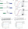
Figure 1.
Figure 1.
TtCas6A and TtCas6B both cleave repeats R1 and R3 and retain their cleaved products. (A) Sequences and predicted secondary structures of T. thermophilus CRISPR repeats. Sites of cleavage are indicated with blue arrows. TtCas6e (TTHB192) cleaves repeat R2, while TtCas6A (TTHA0078) and TtCas6B (TTHB231) both cleave repeats R1 and R3. (B) Cleavage product binding affinities of TtCas6A and TtCas6B enzymes. Maltose-binding protein (MBP)-fused TtCas6A or TtCas6B were bound to 5′-[32P]-radiolabeled, in vitro transcribed R1 and R3 RNAs. Bound and unbound fractions were resolved by electrophoresis on a native polyacrylamide gel and visualized by phosphorimaging. The data for these and all subsequent binding assays were fit with standard binding isotherms (solid line), unless otherwise stated. Error bars on each data point denote standard error of the mean (SEM) from three independent experiments. (C) Kinetics experiments to confirm single turnover. RNA cleavage assays were carried out at indicated protein:RNA ratios. RNA cleavage was monitored using denaturing polyacrylamide gel electrophoresis. The data from these and all subsequent endoribonuclease activity assays were fit with single exponential curves to yield first-order rate constants.
Figure 2.
Figure 2.
Structures of TtCas6A and TtCas6B enzymes bound to substrate mimic and product RNAs. (A) Ribbon diagrams showing the overall views of Cas6–RNA complexes: TtCas6A–R1 substrate mimic (left), TtCas6A–R1 product (middle) and TtCas6B–R3 product (right). Bound RNAs are depicted in cartoon format and colored in yellow. The scissile phosphate groups are depicted as orange spheres. All cartoon molecular diagrams were generated using Pymol (http://www.pymol.org). (B) Zoomed-in views of the TtCas6 active sites, shown in the same orientation as in A. Hydrogen-bonding interactions are denoted with dashed lines; numbers indicate interatomic distances in Å. (C) Endonuclease activity assays of wild-type (WT) and active-site mutant proteins. For the TtCas6A H37A mutant, the cleavage assay was additionally carried out in the presence of 500 mM imidazole. (D) Active site of TtCas6A undergoes conformational ordering on substrate recognition. Left: zoomed-in view of the active site in the RNA-free TtCas6A molecule in the 2:1 protein–R1 substrate mimic complex. Right: zoomed-in view of the active site in the RNA-bound TtCas6A molecule. Hydrogen-bonding interactions are denoted with dashed lines.
Figure 3.
Figure 3.
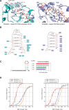
RNA recognition by TtCas6A and TtCas6B. (A) Detailed views of RNA binding by TtCas6A (left) and TtCas6B (right). Hydrogen-bonding interactions are indicated with black dashed lines. Blue spheres denote backbone amide nitrogen atoms of Lys226 in TtCas6A (left) and Ala145 and Lys253 in TtCas6B (right). (B) Schematic diagrams of protein–RNA contacts in the TtCas6A–R1 substrate mimic (left) and TtCas6B–R3 product complexes. Amino acid residues contacting the bound RNA via ionic or hydrogen-bonding interactions are highlighted. Blue arrows mark the scissile phosphates. Red circles denote phosphodiester groups in the RNA backbone. Red lines indicate base-pairing interactions. (C) Base-pair contributions to R1 repeat recognition by TtCas6A. A series of RNAs in which individual C-G base pairs were substituted with A-U were prepared and assayed for binding to TtCas6A using EMSAs. The data for each base-pair substitution are expressed as Kd and as fold reduction in affinity relative to wild-type R1 RNA. The color-coding follows the schematic diagram of the R1 RNA (left). (D) R1 (left) or R3 (right) product RNA binding by WT TtCas6A, R129A or H37A mutants was quantified using EMSAs. The data are plotted as in Figure 1B, with the exception of TtCas6A H37A, for which a modified equation using a Hill coefficient for negative cooperativity (n = 0.6) was used.
Figure 4.
Figure 4.
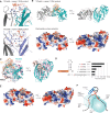
Recognition of the 5′ segment of the repeat RNA. (A) Details of sequence-specific recognition of nucleotides upstream of the stem-loop in RNA repeats. Top: TtCas6A–R1 product complex. Nucleotides 1–14 of the R1 product RNA are disordered. Bottom: TtCas6B–R3 product complex. Nucleotides 1–15 of the R3 product RNA are disordered. (B) Surface electrostatic potential map of TtCas6A identifies a second RNA binding site. Top: Cartoon diagram of the 2:1 TtCas6A–R1 product RNA complex. RNA is shown in orange. Bound sulfate ions are depicted in stick format. Bottom: Electrostatic surface potential map of TtCas6A, shown in the same orientations as earlier. Blue, positively charged region; red, negatively charged region. The positively charged patch located on the surface opposite from the active site is highlighted with a black ellipse. (C) Structural superposition of the TtCas6A–R1 product RNA (TtR1) and PfCas6–repeat RNA (PfRNA) (PDB code: 3PKM) complexes. TtCas6A is colored teal; PfCas6 is colored pink. T. thermophilus R1 repeat RNA is colored orange. PfRNA is colored black. Nucleotide A15 of TtR1 aligns with G10 of PfRNA. (D) Nucleotides in the single-stranded 5′ segment of R1 repeat RNA contribute to binding. TtCas6A binding to a series of truncated RNAs based on the R1 repeat was quantified by EMSAs as in Figure 1B. The data are expressed as Kd and as a fold binding defect relative to wild-type R1 repeat. 5′-terminal G nucleotides resulting from in vitro transcription are shown in G. (E) Structural superposition of TtCas6A dimer with P. furiosus repeat RNA, based on the superposition shown in D. TtCas6A is colored according to surface electrostatic potential and shown in the same orientations as in B. TtR1 RNA is colored orange; PfRNA is colored black. (F) Cartoon model of RNA recognition by TtCas6 enzymes. TtCas6A binds the stem-loop region of the RNA (red solid line) at the interface of the two RRM-like domains. The two major elements responsible for the interaction are the variable beta-hairpin and the Gly-rich loop (both depicted in blue). Additionally, the 5′ segment of the repeat RNA (dashed red line) is bound by a distal positively charged cleft.

Similar articles

Cited by

References

    1. Wiedenheft B, Sternberg SH, Doudna JA. RNA-guided genetic silencing systems in bacteria and archaea. Nature. 2012;482:331–338. - PubMed
    1. Marraffini LA, Sontheimer EJ. CRISPR interference: RNA-directed adaptive immunity in bacteria and archaea. Nat. Rev. Genet. 2010;11:181–190. - PMC - PubMed
    1. Al-Attar S, Westra ER, van der Oost J, Brouns SJJ. Clustered regularly interspaced short palindromic repeats (CRISPRs): the hallmark of an ingenious antiviral defense mechanism in prokaryotes. Biol. Chem. 2011;392:277–289. - PubMed
    1. Terns MP, Terns RM. CRISPR-based adaptive immune systems. Curr. Opin. Microbiol. 2011;14:321–327. - PMC - PubMed
    1. Brouns SJJ, Jore MM, Lundgren M, Westra ER, Slijkhuis RJH, Snijders APL, Dickman MJ, Makarova KS, Koonin EV, van der Oost J. Small CRISPR RNAs guide antiviral defense in prokaryotes. Science. 2008;321:960–964. - PMC - PubMed

Publication types

MeSH terms