

Residual fMRI sensitivity for identity changes in acquired prosopagnosia
- PMID: 24151479
- PMCID: PMC3799008
- DOI: 10.3389/fpsyg.2013.00756
Residual fMRI sensitivity for identity changes in acquired prosopagnosia
Abstract
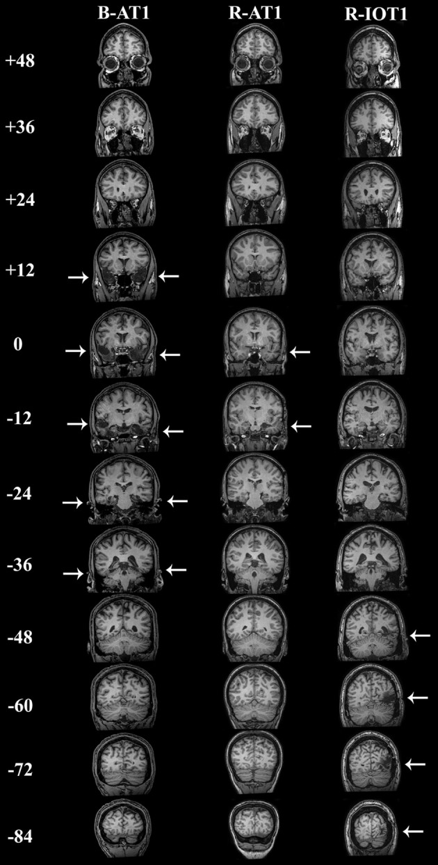
While a network of cortical regions contribute to face processing, the lesions in acquired prosopagnosia are highly variable, and likely result in different combinations of spared and affected regions of this network. To assess the residual functional sensitivities of spared regions in prosopagnosia, we designed a rapid event-related functional magnetic resonance imaging (fMRI) experiment that included pairs of faces with same or different identities and same or different expressions. By measuring the release from adaptation to these facial changes we determined the residual sensitivity of face-selective regions-of-interest. We tested three patients with acquired prosopagnosia, and all three of these patients demonstrated residual sensitivity for facial identity changes in surviving fusiform and occipital face areas of either the right or left hemisphere, but not in the right posterior superior temporal sulcus. The patients also showed some residual capabilities for facial discrimination with normal performance on the Benton Facial Recognition Test, but impaired performance on more complex tasks of facial discrimination. We conclude that fMRI can demonstrate residual processing of facial identity in acquired prosopagnosia, that this adaptation can occur in the same structures that show similar processing in healthy subjects, and further, that this adaptation may be related to behavioral indices of face perception.
Keywords: adaptation; expression; fMRI; face perception; identity; prosopagnosia; sensitivity.
Figures


Similar articles
-
Understanding the functional neuroanatomy of acquired prosopagnosia.Neuroimage. 2007 Apr 1;35(2):836-52. doi: 10.1016/j.neuroimage.2006.09.051. Epub 2007 Feb 14. Neuroimage. 2007. PMID: 17303440
-
The roles of "face" and "non-face" areas during individual face perception: evidence by fMRI adaptation in a brain-damaged prosopagnosic patient.Neuroimage. 2008 Mar 1;40(1):318-32. doi: 10.1016/j.neuroimage.2007.11.012. Epub 2007 Nov 22. Neuroimage. 2008. PMID: 18164628
-
Reconstructing dynamic mental models of facial expressions in prosopagnosia reveals distinct representations for identity and expression.Cortex. 2015 Apr;65:50-64. doi: 10.1016/j.cortex.2014.11.015. Epub 2014 Dec 24. Cortex. 2015. PMID: 25638352
-
Face processing in the temporal lobe.Handb Clin Neurol. 2022;187:191-210. doi: 10.1016/B978-0-12-823493-8.00019-5. Handb Clin Neurol. 2022. PMID: 35964972 Review.
-
[Prosopagnosia and facial expression recognition].Brain Nerve. 2014 Apr;66(4):393-402. Brain Nerve. 2014. PMID: 24748087 Review. Japanese.
Cited by
-
Voice Recognition in Face-Blind Patients.Cereb Cortex. 2016 Apr;26(4):1473-1487. doi: 10.1093/cercor/bhu240. Epub 2014 Oct 27. Cereb Cortex. 2016. PMID: 25349193 Free PMC article.
-
Interrogating an ICD-coded electronic health records database to characterize the epidemiology of prosopagnosia.J Clin Transl Sci. 2020 Jun 19;5(1):e11. doi: 10.1017/cts.2020.497. J Clin Transl Sci. 2020. PMID: 33948237 Free PMC article.
-
The Face-Processing Network Is Resilient to Focal Resection of Human Visual Cortex.J Neurosci. 2016 Aug 10;36(32):8425-40. doi: 10.1523/JNEUROSCI.4509-15.2016. J Neurosci. 2016. PMID: 27511014 Free PMC article.
-
Dissociable roles of the inferior longitudinal fasciculus and fornix in face and place perception.Elife. 2015 Aug 29;4:e07902. doi: 10.7554/eLife.07902. Elife. 2015. PMID: 26319355 Free PMC article.
-
Aftereffects in face processing.Front Psychol. 2013 Nov 14;4:854. doi: 10.3389/fpsyg.2013.00854. eCollection 2013. Front Psychol. 2013. PMID: 24294209 Free PMC article. No abstract available.
References
Grants and funding
LinkOut - more resources
Full Text Sources
Other Literature Sources
Molecular Biology Databases