. 2013 Oct 1;6(5):562-72.
doi: 10.1593/tlo.13409. eCollection 2013.

The Effect of Molecular Weight, PK, and Valency on Tumor Biodistribution and Efficacy of Antibody-Based Drugs

Affiliations

The Effect of Molecular Weight, PK, and Valency on Tumor Biodistribution and Efficacy of Antibody-Based Drugs

Ruth Muchekehu et al. Transl Oncol. .

Abstract

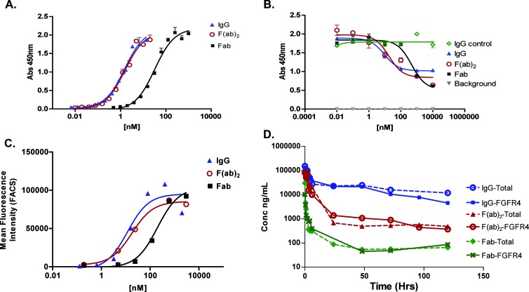
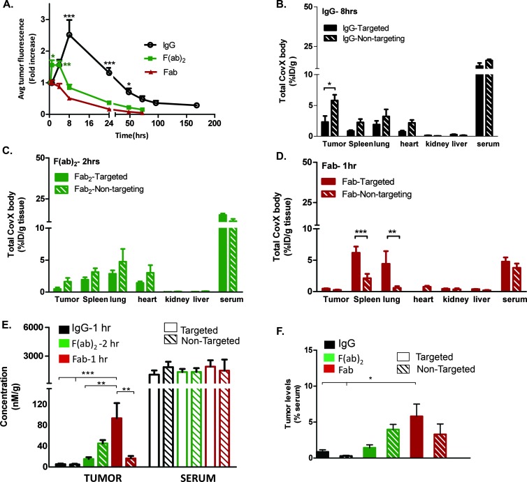
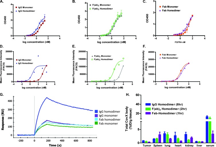
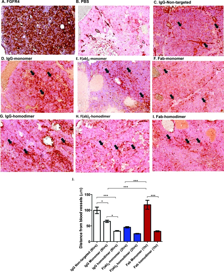
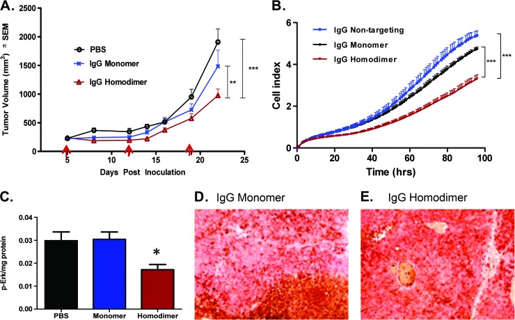
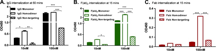
Poor drug delivery and penetration of antibody-mediated therapies pose significant obstacles to effective treatment of solid tumors. This study explored the role of pharmacokinetics, valency, and molecular weight in maximizing drug delivery. Biodistribution of a fibroblast growth factor receptor 4 (FGFR4) targeting CovX-body (an FGFR4-binding peptide covalently linked to a nontargeting IgG scaffold; 150 kDa) and enzymatically generated FGFR4 targeting F(ab)2 (100 kDa) and Fab (50 kDa) fragments was measured. Peak tumor levels were achieved in 1 to 2 hours for Fab and F(ab)2 versus 8 hours for IgG, and the percentage injected dose in tumors was 0.45%, 0.5%, and 2.5%, respectively, compared to 0.3%, 2%, and 6% of their nontargeting controls. To explore the contribution of multivalent binding, homodimeric peptides were conjugated to the different sized scaffolds, creating FGFR4 targeting IgG and F(ab)2 with four peptides and Fab with two peptides. Increased valency resulted in an increase in cell surface binding of the bivalent constructs. There was an inverse relationship between valency and intratumoral drug concentration, consistent with targeted consumption. Immunohistochemical analysis demonstrated increased size and increased cell binding decreased tumor penetration. The binding site barrier hypothesis suggests that limited tumor penetration, as a result of high-affinity binding, could result in decreased efficacy. In our studies, increased target binding translated into superior efficacy of the IgG instead, because of superior inhibition of FGFR4 proliferation pathways and dosing through the binding site barrier. Increasing valency is therefore an effective way to increase the efficacy of antibody-based drugs.


Figures

Figure 1
Figure 1
Characterization of FGFR4 binding scaffolds. (A) IgG, F(ab)2, and Fab constructs bind specifically to FGFR4. (B) In an FGF19 competition ELISA, all constructs compete with FGF19 to bind to FGFR4. (C) All constructs bind to Huh-7 cells. (D) PK curves of IgG, F(ab)2, and Fab following a single i.v. dose of 10 mg/kg in Swiss Webster mice. Both total and FGFR4 binding were measured as described in the Materials and Methods section.
Figure 2
Figure 2
Biodistribution studies. (A) Time-dependent tumor uptake of IgG, F(ab)2, and Fab. In vivo optical imaging of near infra-red (NIR)-conjugated constructs. Average signal intensities were quantified using regions of interest (ROIs) from the tumor sites. Data are presented as mean fold increase from initial image capture at 30 minutes ± SEM of eight mice (***P < .001, *P < .05; IgG vs both F(ab)2 and Fab accumulation, *P < .05 and **P < .01; F(ab)2 vs Fab accumulation by two-way ANOVA with Bonferroni post-test). Tumor and normal tissue uptake of (B) IgG 8 hours post dose (*P < .05), (C) F(ab)2 2 hours post dose, and (D) Fab 1 hour post dose (***P < .001, **P < .01 by two-way ANOVA with Bonferroni post-test). (E) IgG, F(ab)2, and Fab tumor uptakes and serum levels compared at the early time points of 1 hour, 2 hours, and 1 hour, respectively. At this early time point with equivalent serum levels, the targeted Fab shows maximal accumulation levels compared to the F(ab)2 and IgG (***P < .001, **P < .01 by one-way ANOVA with Bonferroni post-test). (F) Tumor to serum levels further demonstrate that the Fab construct accumulation is significantly higher than the IgG accumulation (*P < .05 by two-way ANOVA with Bonferroni post-test).
Figure 3
Figure 3
Increasing valency increases cell binding of bivalent constructs. (A–C) Monomer and homodimer peptide-conjugated constructs bind recombinant FGFR4 in a similar manner on an FGFR4 binding ELISA. Huh-7 cell binding of IgG (D) and F(ab)2 (E) homodimer-conjugated constructs demonstrates a boost in cell binding affinity, whereas the Fab monomer and homodimer (F) have similar cell binding affinities. (G) Anti-idiotype capture levels of anti-FGFR4 monomer and homodimer compounds to measure multivalent interactions. Increased levels of FGFR4 binding is seen with the IgG homodimer. (H) Tumor and normal tissue uptake of homodimer peptide-conjugated IgG, F(ab)2, and Fab (*P < .05 vs monomer IgG).
Figure 4
Figure 4
Increased avidity decreases penetration of scaffolds into the tumor. (A) FGFR4 staining in an adjacent section is shown. Dual staining for blood vessels (brick red) and human IgG (brown) is shown in (B) PBS-dosed animals, (C) nontargeted IgG (8 hours), monomer peptide-conjugated (D) IgG (8 hours), (E) F(ab)2 (2 hours), (F) Fab (1 hour) and homodimer peptide-conjugated (G) IgG (8 hours), (H) F(ab)2 (2 hours) and (I) Fab (1 hour). Arrows indicate blood vessels (red). Perivascular or diffuse construct staining from those points can be seen. (J) Plot of average distance from randomly selected blood vessels (mean ± SEM; *P < .05, ***P < .001 by one-way ANOVA with Bonferroni post-test).
Figure 5
Figure 5
Increased avidity leads to superior efficacy. (A) In vivo xenograft study (i.p. dosing, 30 mg/kg once weekly) and (B) in vitro cell proliferation assay (12 nM compounds present in media for whole experiment). *P < .05, ***P < .001 by two-way ANOVA with Bonferroni post-test. Arrows indicate dosing. (C) Phospho-Erk levels measured in tumors harvested at the end of the efficacy study; *P < .05 versus PBS-dosed tumors by two-tailed Student's t test. Dual staining of blood vessels and human IgG of the tumors at the end of the efficacy study; (D) monomer peptide IgG dosed and (E) homodimer peptide IgG dosed.
Figure 6
Figure 6
Multivalent binding is required for internalization. Internalization assays were conducted by incubating Huh-7 cells with biotinylated constructs at concentrations indicated at 37°C. Maximal internalization was seen at (A) 60 minutes for IgG constructs and 15 minutes for the (B) F(ab)2 and (C) Fab constructs. The average ± SD is shown (*P < .05, **P < .01, ***P < .001 by two-way ANOVA with Bonferroni posttest).

References

    1. Tunggal JK, Cowan DS, Shaikh H, Tannock IF. Penetration of anticancer drugs through solid tissue: a factor that limits the effectiveness of chemotherapy for solid tumors. Clin Cancer Res. 1999;5:1583–1586. - PubMed
    1. Sedlacek HH, Seemann G, Hoffmann D, Czech J, Lorenz P, Kolar C, Bosslet K. Antibodies as carriers of cytotoxicity. In: Huber H, Queiber W, editors. Contributions to Oncology. Vol. 43. Basel, Switzerland: Karger; 1992. pp. 74–75.
    1. Ackerman ME, Pawlowski D, Wittrup KD. Effect of antigen turnover rate and expression level on antibody penetration into tumor spheroids. Mol Cancer Ther. 2008;7:2233–2240. - PMC - PubMed
    1. Chaplin DJ, Olive PL, Durand RE. Intermittent blood flow in a murine tumor: radiobiological effects. Cancer Res. 1987;47:597–601. - PubMed
    1. Heldin CH, Rubin K, Pietras K, Ostman A. High interstitial fluid pressure—an obstacle in cancer therapy. Nat Rev Cancer. 2004;4:806–813. - PubMed