. 2013 Oct 1;6(5):596-606.
doi: 10.1593/tlo.13412. eCollection 2013.

Colon Macrophages Polarized by Commensal Bacteria Cause Colitis and Cancer through the Bystander Effect

Affiliations

Colon Macrophages Polarized by Commensal Bacteria Cause Colitis and Cancer through the Bystander Effect

Yonghong Yang et al. Transl Oncol. .

Abstract

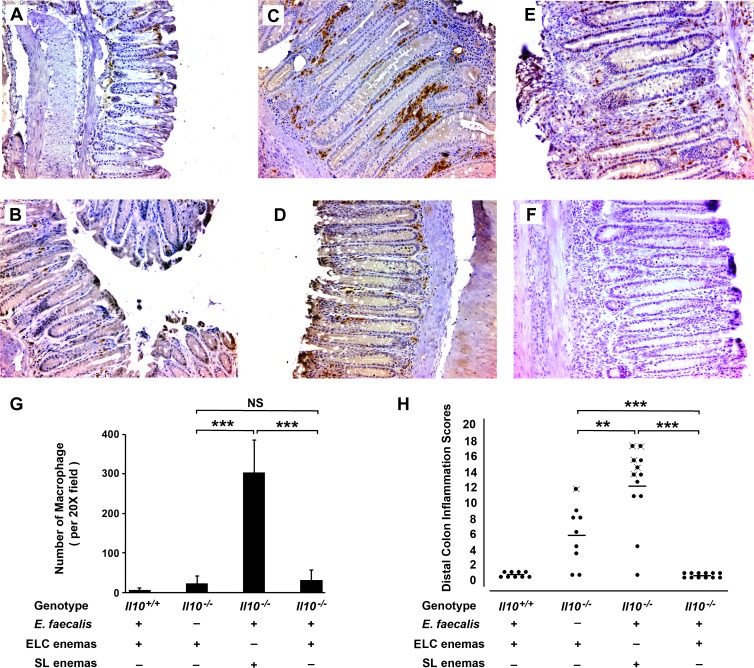
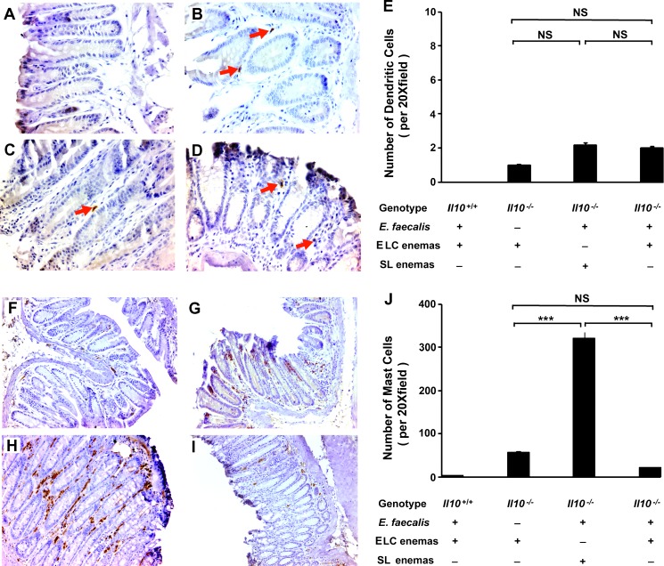
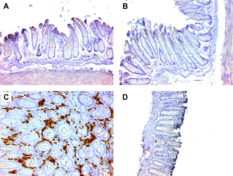
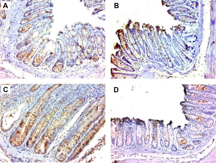
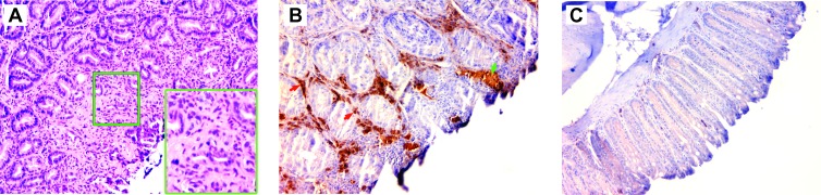
Intestinal commensal bacteria have recently been shown to trigger macrophages to produce diffusible clastogens (or chromosome-breaking factors) through a bystander effect (BSE) that mediates DNA damage and induces chromosomal instability in neighboring cells. Colon macrophages appear central to colon carcinogenesis and BSE through the expression of tumor necrosis factor-α (TNF-α) and cyclooxygenase-2 (COX-2). The former induces netrin-1, a regulator of intestinal epithelial cell apoptosis, and the latter generates trans-4-hydroxy-2-nonenal (4-HNE), an endogenous mutagen. To test whether colon macrophages are key effectors for BSE, we depleted these cells in interleukin-10 knockout mice colonized with Enterococcus faecalis using encapsulated liposomal clodronate (ELC), a bisphosphonate that causes macrophage apoptosis. We observed that E. faecalis polarizes colon macrophages to an M1 phenotype. In addition, depleting these cells suppressed COX-2 and TNF-α, blocked the formation of 4-HNE protein adducts, and inhibited up-regulation of netrin-1-all markers for BSE. Finally, treatment with ELC prevented colitis, β-catenin activation, and cancer formation. These results show that selected human commensals can polarize colon macrophages to the M1 phenotype and, when activated, serve as the key effector for bacterial-induced BSE. Our findings suggest that depleting M1-polarized macro-phages is a mechanism for the chemopreventive activity of bisphosphonates and that it represents a new strategy for preventing colon cancer induced by intestinal commensals.


Figures

Figure 1
Figure 1
Depleting colon macrophages with ELC suppresses inflammation and prevents cancer in Il10-/- mice colonized with E. faecalis. (A) Immunohistochemical staining of colon biopsies using anti-F4/80 antibody (brown) shows few cells in the lamina propria of wild-type mice (i.e., Il10+/+) colonized with E. faecalis and administered ELC (n = 9). (B) Staining of sham-colonized Il10-/- mice administered ELC also show few macrophages (n = 9). (C) Numerous macrophages are present in colon biopsies from E. faecalis-colonized Il10-/- mice administered SL (n = 12). (D) Macrophage staining is infrequent in E. faecalis-colonized Il10-/- mice administered ELC (n = 12). (E) Macro-phages in the lamina propria of colon biopsies from E. faecalis-colonized Il10-/- mice administered SL are positive for iNOS, suggesting that E. faecalis polarized these cells to an M1 phenotype. (F) Staining of biopsies in E for arginase is negative and consistent with the M1 polarization of macrophages. (G) Colon macrophages for mice administered ELC are significantly lower than for mice given SL (***P < .001). (H) Inflammatory scores are significantly lower for colon biopsies from E. faecalis-colonized and sham-colonized Il10-/- mice administered ELC compared to SL-treated mice (***P < .001 and **P < .01, respectively); among ELC-treated mice, inflammation scores are higher for sham-colonized Il10-/- mice compared to E. faecalis-colonized mice (***P < .001); more colon cancers are seen in SL-treated and E. faecalis-colonized mice than similarly colonized and ELC-treated mice (mice with cancer indicated by crossed closed circles; P <.05). All photomicrographs are x20.
Figure 2
Figure 2
Effect of ELC treatment on colonic dendritic cells and mast cells. (A) Immunohistochemical staining of colon biopsies for mouse dendritic cell marker (33D1) is negative for wild-type mice colonized with E. faecalis and administered ELC (n = 9). (B–D) Few 33D1-positive cells are seen in colon biopsies for Il10-/- mice (red arrows). (E) Average number of 33D1-positive cells per x20 field across Il10-/- groups (NS, not significant). (F) Immunohistochemical staining of colon biopsies for mast cell tryptase shows few cells from biopsies of wild-type mice colonized with E. faecalis and administered ELC. (G) Increased mast cell staining is seen in biopsies for sham-colonized Il10-/- mice administered ELC (n = 9). (H) Larger increase in mast cell staining is noted in biopsies for E. faecalis-colonized Il10-/- mice administered SL (n = 12). (I) Depleting macrophages with ELC decreases mast cell staining in biopsies for E. faecalis-colonized Il10-/- mice (n = 12). (J) Average number of mast cells per x20 field across groups (NS, not significant; ***P < .001). All photomicrographs are x20.
Figure 3
Figure 3
Depleting colon macrophages is associated with reduced COX-2 expression. (A) Immunohistochemical staining shows no COX-2 in colon biopsies in wild-type mice colonized with E. faecalis and administered ELC. (B) COX-2 expression is noted for sham-colonized Il10-/- mice administered ELC. (C) Increased COX-2 expression is seen for Il10-/- mice colonized with E. faecalis and administered SL. (D) Depleting macrophages with ELC significantly decreases COX-2 expression for E. faecalis-colonized Il10-/- mice. All photomicrographs are x20.
Figure 4
Figure 4
Depleting colon macrophages decreases 4-HNE protein adducts. (A) Immunohistochemical staining for 4-HNE protein adducts was negative in colon biopsies from wild-type mice colonized with E. faecalis and administered ELC. (B) Slight staining is noted for 4-HNE protein adducts in the lamina propria and epithelium for sham-colonized Il10-/- mice administered ELC. (C) Colon crypts (red arrow) and lamina propria (green arrow) stain strongly for 4-HNE protein adducts for Il10-/- mice colonized with E. faecalis and administered SL. (D) Staining for 4-HNE protein adducts is reduced for E. faecalis-colonized Il10-/- mice administered ELC. All photomicrographs are x20.
Figure 5
Figure 5
Depleting colon macrophages inhibits TNF-α production. (A) Immunohistochemical staining for TNF-α-positive cells is not evident in colon biopsies from wild-type mice colonized with E. faecalis and administered ELC. (B) A few TNF-α-positive cells are noted in the lamina propria of biopsies from sham-colonized Il10-/- mice administered ELC. (C) In contrast, numerous cells stain strongly for TNF-α for E. faecalis-colonized Il10-/- mice administered SL. (D) Few TNF-α-positive cells are evident in the lamina propria of colons from E. faecalis-colonized Il10-/- mice administered ELC. All photomicrographs are x20.
Figure 6
Figure 6
Depleting colon macrophages blocks TNF-α-induced netrin-1 expression. (A) Typical expression of netrin-1 (brown) in epithelial cells is observed at the bottom of colon crypts for wild-type mice colonized with E. faecalis and administered ELC. (B) Similar staining is noted for netrin-1 staining for sham-colonized Il10-/- mice administered ELC. (C) Colonic epithelial cells stain more strongly for netrin-1 in biopsies from E. faecalis-colonized Il10-/- mice administered SL. (D) ELC decreases netrin-1 staining in biopsies from E. faecalis-colonized Il10-/- mice to levels similar to that found in wild-type mice colonized with E. faecalis. All photomicrographs at x20.
Figure 7
Figure 7
Depleting colon macrophages inhibits Wnt/β-catenin signaling. (A) Hematoxylin and eosin staining shows typical cancer in distal colon of an E. faecalis-colonized Il10-/- mouse administered SL at x20 and x40 (inset). (B) Immunohistochemical staining for unphosphorylated (active) β-catenin shows strong signaling in neoplastic epithelial cells (green arrow) and inflammatory cells in the lamina propria (red arrows) for Il10-/- mice colonized with E. faecalis and administered SL (x20 magnification). (C) No active β-catenin is evident in biopsies from E. faecalis-colonized Il10-/- mice administered ELC (x20).

References

    1. Grivennikov SI, Greten FR, Karin M. Immunity, inflammation, and cancer. Cell. 2010;140:883–899. - PMC - PubMed
    1. Colotta F, Allavena P, Sica A, Garlanda C, Mantovani A. Cancer-related inflammation, the seventh hallmark of cancer: links to genetic instability. Carcinogenesis. 2009;30:1073–1081. - PubMed
    1. Qian BZ, Pollard JW. Macrophage diversity enhances tumor progression and metastasis. Cell. 2010;141:39–51. - PMC - PubMed
    1. Gounaris E, Erdman SE, Restaino C, Gurish MF, Friend DS, Gounari F, Lee DM, Zhang G, Glickman JN, Shin K, et al. Mast cells are an essential hematopoietic component for polyp development. Proc Natl Acad Sci USA. 2007;104:19977–19982. - PMC - PubMed
    1. Wu S, Rhee KJ, Albesiano E, Rabizadeh S, Wu X, Yen HR, Huso DL, Brancati FL, Wick E, McAllister F, et al. A human colonic commensal promotes colon tumorigenesis via activation of T helper type 17 T cell responses. Nat Med. 2009;15:1016–1022. - PMC - PubMed