. 2013:2013:107238.
doi: 10.1155/2013/107238. Epub 2013 Sep 15.

Extending the serum half-life of G-CSF via fusion with the domain III of human serum albumin

Affiliations

Extending the serum half-life of G-CSF via fusion with the domain III of human serum albumin

Shuqiang Zhao et al. Biomed Res Int. 2013.

Abstract

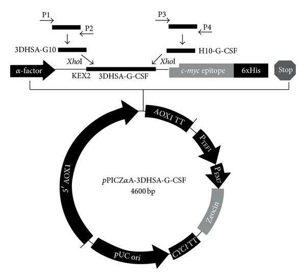
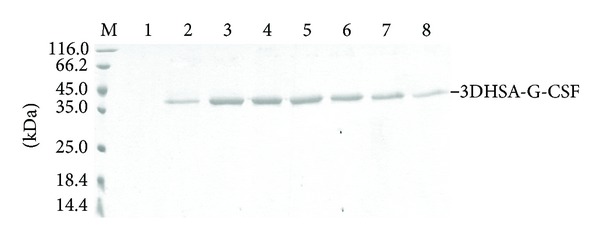
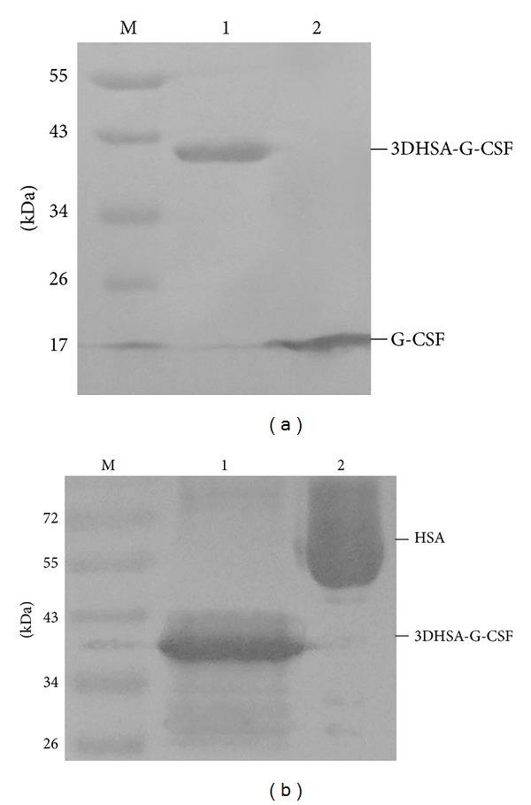
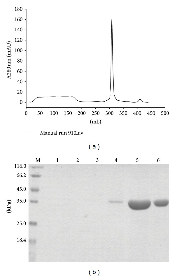
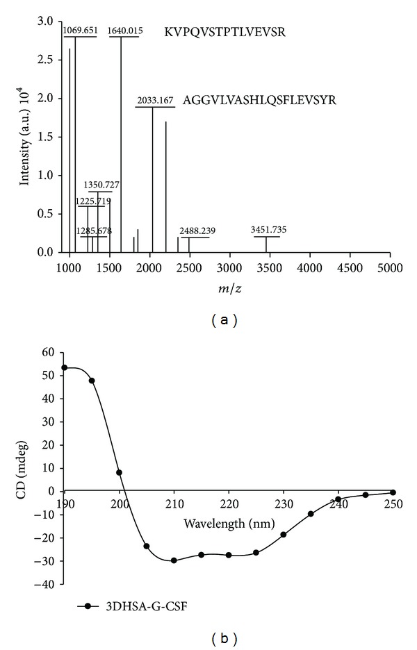
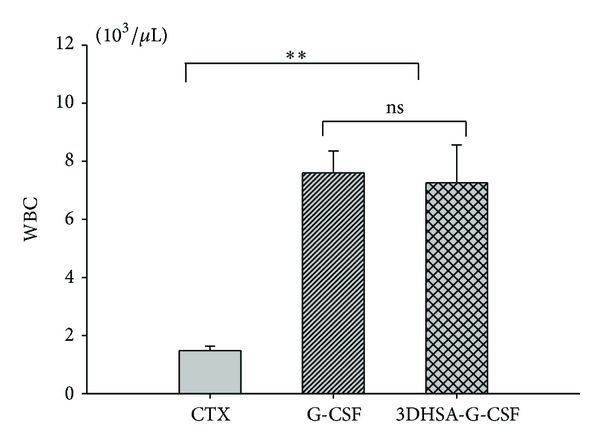
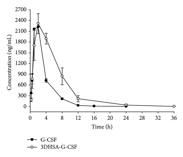
Protein fusion technology is one of the most commonly used methods to extend the half-life of therapeutic proteins. In this study, in order to prolong the half-life of Granulocyte colony stimulating factor (G-CSF), the domain III of human serum albumin (3DHSA) was genetically fused to the N-terminal of G-CSF. The 3DHSA-G-CSF fusion gene was cloned into pPICZ α A along with the open reading frame of the α -factor signal under the control of the AOX1 promoter. The recombinant expression vector was transformed into Pichia pastoris GS115, and the recombinant strains were screened by SDS-PAGE. As expected, the 3DHSA-G-CSF showed high binding affinity with HSA antibody and G-CSF antibody, and the natural N-terminal of 3DHSA was detected by N-terminal sequencing. The bioactivity and pharmacokinetic studies of 3DHSA-G-CSF were respectively determined using neutropenia model mice and human G-CSF ELISA kit. The results demonstrated that 3DHSA-G-CSF has the ability to increase the peripheral white blood cell (WBC) counts of neutropenia model mice, and the half-life of 3DHSA-G-CSF is longer than that of native G-CSF. In conclusion, 3DHSA can be used to extend the half-life of G-CSF.


Figures

Figure 1
Figure 1
Construction of recombinant pPIC-3DHSA-G-CSF eukaryotic expression vector. The synthesized HSA and G-CSF genes were used as templates for the amplification of 3DHSA-G10 and H10-G-CSF genes by PCR with primer 1 and primer 2, primer 3 and primer 4, respectively. The purified 3DHSA-G10 and H10-G-CSF genes were used as templates for obtaining 3DHSA-G-CSF gene by overlap PCR. Both pPICZαA plasmid and 3DHSA-G-CSF gene were digested by XhoI and ligated together by T4 DNA ligase to construct expression vector pPIC-3DHSA-G-CSF. 3DHSA-G-CSF was inserted into the sites XhoI of pPICZαA along with the open reading frame of the α-factor signal under the control of the AOX1 promoter. The KEX2 cleavage site was located between the signal peptide and the 3DHSA-G-CSF sequences.
Figure 2
Figure 2
Analysis of transformants that expressed 3DHSA-G-CSF by SDS-PAGE. The culture supernatants of screened transformant were resolved by 12% SDS-PAGE and stained with Coomassie Blue. M: protein marker; Lanes 1–8 correspond to different transformants of GS115-pPIC-3DHSA-G-CSF; the protein band about 42 kDa was marked 3DHSA-G-CSF on the right of SDS-PAGE.
Figure 3
Figure 3
Western blotting analysis using primary antibody anti-G-CSF and anti-HSA. M: prestained protein marker, (a) antibody anti-G-CSF, Lane a1: 3DHSA-G-CSF, Lane a2: G-CSF; (b) antibody anti-HSA, Lane b1: 3DHSA-G-CSF, Lane b2: HSA.
Figure 4
Figure 4
Purification of 3DHSA-G-CSF. The supernatants containing the target proteins were purified by Cibacron Blue F3G-A Sepharose and Butyl-Sepharose 4B column, and the final purity was detected by 12% SDS-PAGE. (a) The hydrophobic chromatograph of 3DHSA-G-CSF by Butyl-Sepharose 4B column. (b) 12% SDS-PAGE, M: protein marker, Lanes 1~3: flow through, Lanes 4~6: elute.
Figure 5
Figure 5
Structural analysis of purified fusion protein by peptide mass mapping and circular dichroism spectrum. (a) Peptide mass mapping of 3DHSA-G-CSF, (b) circular dichroism spectra of 3DHSA-G-CSF.
Figure 6
Figure 6
In vivo bioactivity of the purified 3DHSA-G-CSF. Both G-CSF and 3DHSA-G-CSF could increase the white blood cell counts in a cyclophosphamide-induced neutropenia model murine, and more potent stimulate was observed compared with CTX (P < 0.01). G-CSF versus 3DHSA-G-CSF not significant (ns) (P > 0.05). Values are expressed as mean ± SD among six samples from one experiment. Data are representative of three independent experiments with similar result.
Figure 7
Figure 7
G-CSF level in serum after subcutaneous (S.C.) administration of G-CSF and 3DHSA-G-CSF. The concentration-time curve of 3DHSA-G-CSF is superior than that of G-CSF. Solid black circle means G-CSF. Black hollow circle means 3DHSA-G-CSF. Each point represents the mean value, and error bars represent SD of the mean (n = 3). Data are representative of three independent experiments with similar result.

References

    1. Nagata S, Tsuchiya M, Asano S. Molecular cloning and expression of cDNA for human granulocyte colony-stimulating factor. Nature. 1986;319(6052):415–418. - PubMed
    1. Nomura H, Imazeki I, Oheda M, et al. Purification and characterization of human granulocyte colony-stimulating factor (G-CSF) The EMBO journal. 1986;5(5):871–876. - PMC - PubMed
    1. Sheikh H, Colaco R, Lorigan P, et al. Use of G-CSF during concurrent chemotherapy and thoracic radiotherapy in patients with limited-stage small-cell lung cancer safety data from a phase II trial. Lung Cancer. 2011;74(1):75–79. - PubMed
    1. Meuer K, Pitzer C, Teismann P, et al. Granulocyte-colony stimulating factor is neuroprotective in a model of Parkinson’s disease. Journal of Neurochemistry. 2006;97(3):675–686. - PubMed
    1. Sanchez-Ramos J, Song S, Sava V, et al. Granulocyte colony stimulating factor decreases brain amyloid burden and reverses cognitive impairment in Alzheimer’s mice. Neuroscience. 2009;163(1):55–72. - PMC - PubMed

Publication types