. 2014 Jul;20(7):543-52.
doi: 10.1089/ten.TEC.2013.0370. Epub 2013 Dec 13.

Control of perfusable microvascular network morphology using a multiculture microfluidic system

Affiliations

Control of perfusable microvascular network morphology using a multiculture microfluidic system

Jordan A Whisler et al. Tissue Eng Part C Methods. 2014 Jul.

Abstract

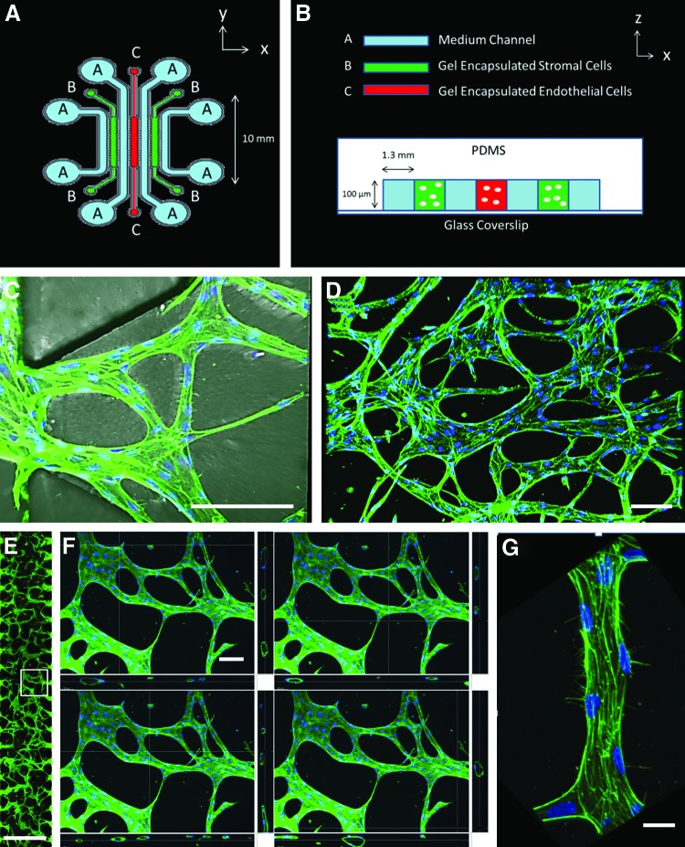
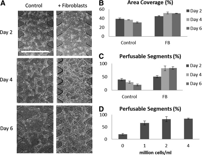
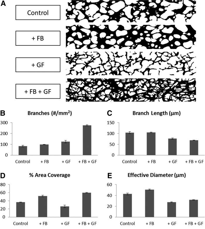
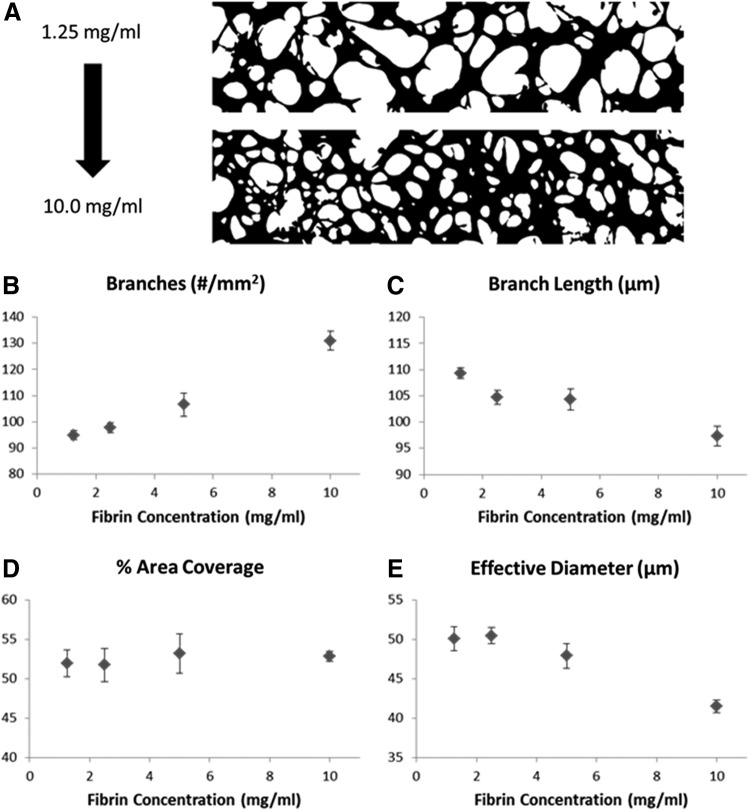
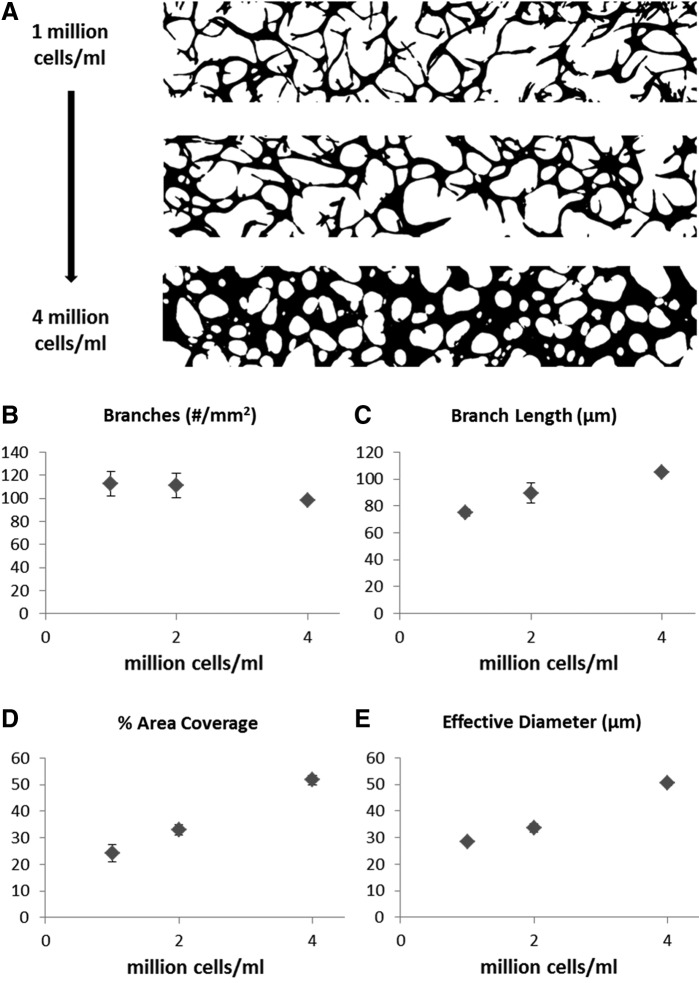
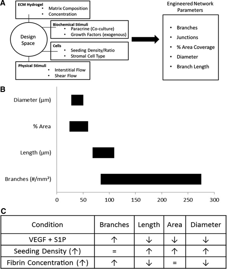
The mechanical and biochemical microenvironment influences the morphological characteristics of microvascular networks (MVNs) formed by endothelial cells (ECs) undergoing the process of vasculogenesis. The objective of this study was to quantify the role of individual factors in determining key network parameters in an effort to construct a set of design principles for engineering vascular networks with prescribed morphologies. To achieve this goal, we developed a multiculture microfluidic platform enabling precise control over paracrine signaling, cell-seeding densities, and hydrogel mechanical properties. Human umbilical vein endothelial cells (HUVECs) were seeded in fibrin gels and cultured alongside human lung fibroblasts (HLFs). The engineered vessels formed in our device contained patent, perfusable lumens. Communication between the two cell types was found to be critical in avoiding network regression and maintaining stable morphology beyond 4 days. The number of branches, average branch length, percent vascularized area, and average vessel diameter were found to depend uniquely on several input parameters. Importantly, multiple inputs were found to control any given output network parameter. For example, the vessel diameter can be decreased either by applying angiogenic growth factors--vascular endothelial growth factor (VEGF) and sphingosine-1-phsophate (S1P)--or by increasing the fibrinogen concentration in the hydrogel. These findings introduce control into the design of MVNs with specified morphological properties for tissue-specific engineering applications.


Figures

<b>FIG. 1.</b>
FIG. 1.
(A) Top view diagram of multiculture microfluidic device containing three parallel gel regions for encapsulation of endothelial and stromal cells. Gel regions (B, C) are separated by medium channels (A) for gas exchange and delivery of nutrients. (B) A cross-sectional view diagram of multiculture microfluidic device. Cell culture region is surrounded by PDMS above and glass coverslip below (not to scale). (C) Top view of perfusable vessels opening to medium channel on left. Region shown is between two PDMS trapezoidal posts. Twenty such regions exist in each microfluidic device. Staining: Green–phalloidin; Blue–DAPI (same for D–G). Scale bar=100 μm. (D) A 10× confocal image of perfusable microvascular network (MVN) grown in a multiculture microfluidic device. Scale bar=100 μm. (E) Full image of vascularized gel region. White box corresponds to enlarged image in (F). Scale bar=1 mm. (F) Sequence of four section views taken throughout 20× confocal stack. Vertical cross sections on the bottom and right correspond to the location of the white crosshairs. As the cross sections progress through the vascularized region, patent lumens are seen to vary in location and diameter, but remain continuous throughout the gel. This demonstrates the existence of perfusable vessels spanning the 100 μm height of the gel region. Further validation of perfusability is provided in the Supplementary videos. Scale bar=100 μm. (G) A 40× confocal image of a segment of perfusable microvessel showing multiple cells. Scale bar=10 μm. See Supplementary video SV1 for three-dimensional rendering with visualization of lumen. Color images available online at www.liebertpub.com/tec
<b>FIG. 2.</b>
FIG. 2.
(A) Phase contrast images (4×) of MVNs formed by endothelial cells (ECs) in isolated monoculture with EGM-2 MV medium (control) or in fibroblast coculture with EGM-2 MV medium. After day 4, control networks regress, while coculture networks remain intact. Scale bar=1 mm. (B) Area covered by MVNs under monoculture and coculture conditions over a 6-day period. Values given are averages over at least three devices with error bars given as standard error (same for C, D). (C) Percentage of perfusable segments as defined by vascularized gel regions opening to medium channel containing at least one patent lumen. (D) Perfusable segments at day 6 as a function of fibroblast seeding density.
<b>FIG. 3.</b>
FIG. 3.
(A) Representative binary images of MVNs formed under conditions of endothelial cell (EC) monoculture in EGM-2 MV medium; EC+Fibroblast (FB) noncontact coculture in EGM-2 MV medium; EC monoculture with EGM-2 MV medium supplemented with vascular endothelial growth factor (VEGF) (50 ng/mL) and sphingosine-1-phosphate (S1P) (250 nM); EC+FB noncontact coculture in EGM-2 MV medium supplemented with VEGF and S1P. Fixed networks were stained with phalloidin and ImageJ was used to process and binarize fluorescent images. One image from each condition was chosen arbitrarily to represent the group. (B) Number of branches per mm2 of vascularized region. Values given are averages over three devices with error bars given as standard error (same for C–E). (C) Average branch length of MVN. (D) Percentage of area covered by perfusable MVN. (E) Effective diameter of vessels in engineered MVN, calculated as the ratio of vascularized area to total length of engineered MVN; GF, growth factor.
<b>FIG. 4.</b>
FIG. 4.
(A) Representative binary images of MVNs formed under conditions of increasing fibrinogen concentration for fibrin gel cell encapsulation experiments. One image from each condition was chosen arbitrarily to represent the group. Fixed networks were stained with phalloidin; ImageJ was used to process and binarize fluorescent images. (B) Number of branches per mm2 of vascularized region. Values given are averages over three to four devices with error bars given as standard error (same for C–E). (C) Average branch length of MVN. (D) Percentage of area covered by perfusable MVN. (E) Effective diameter of vessels in engineered MVN, calculated as the ratio of vascularized area to total length of engineered MVN.
<b>FIG. 5.</b>
FIG. 5.
(A) Representative binary images of MVNs formed under conditions of increasing human umbilical vein endothelial cell (HUVEC) seeding density. One image from each condition was chosen arbitrarily to represent the group. Fixed networks were stained with phalloidin and ImageJ was used to process and binarize fluorescent images. (B) Number of branches per mm2 of vascularized region. Values given are averages over three devices with error bars given as standard error (same for C–E). (C) Average branch length of MVN. (D) Percentage of area covered by perfusable MVN. (E) Effective diameter of vessels in engineered MVN, calculated as the ratio of vascularized area to total length of engineered MVN.
<b>FIG. 6.</b>
FIG. 6.
(A) Diagram of design approach to microvascular tissue engineering. Use of microfluidic platform to quantify effects of mechanical and chemical stimuli on resulting network morphology. Novel, noncontact multiculture system to study cell–cell communication and network stabilization. (B) Range of network parameters achievable through control of inputs studied. (C) Table of the effects of tunable inputs on distinct network morphological parameters.

References

    1. Kassab G.S.Scaling laws of vascular trees: of form and function. Am J Physiol Heart Circ Physiol 290,H894, 2006 - PubMed
    1. Muschler G.F., Nakamoto C., and Griffith L.G.Engineering Principles of Clinical Cell-Based Tissue Engineering. J Bone Jt Surg 86,1541, 2004 - PubMed
    1. Hoganson D.M., et al. Principles of Biomimetic Vascular Network Design Applied to a Tissue-Engineered Liver Scaffold. Tissue Eng Part A 16,1469, 2010 - PMC - PubMed
    1. Murray C.D.The Physiological Principle of Minimum Work I. The Vascular System and the Cost of Blood Volume. Proc Natl Acad Sci 12,207, 1926 - PMC - PubMed
    1. Barber R.W., and Emerson D.R.Biomimetic design of artificial micro-vasculatures for tissue engineering. Altern Lab Anim ATLA 38Suppl 1,67, 2010 - PubMed

Publication types

MeSH terms