

Automated hippocampal segmentation in patients with epilepsy: available free online
- PMID: 24151901
- PMCID: PMC3995014
- DOI: 10.1111/epi.12408
Automated hippocampal segmentation in patients with epilepsy: available free online
Abstract
Purpose: Hippocampal sclerosis, a common cause of refractory focal epilepsy, requires hippocampal volumetry for accurate diagnosis and surgical planning. Manual segmentation is time-consuming and subject to interrater/intrarater variability. Automated algorithms perform poorly in patients with temporal lobe epilepsy. We validate and make freely available online a novel automated method.
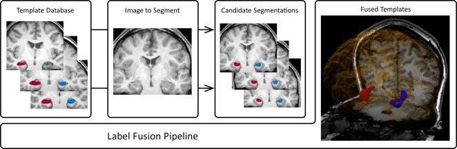
Methods: Manual hippocampal segmentation was performed on 876, 3T MRI scans and 202, 1.5T scans. A template database of 400 high-quality manual segmentations was used to perform automated segmentation of all scans with a multi-atlas-based segmentation propagation method adapted to perform label fusion based on local similarity to ensure accurate segmentation regardless of pathology. Agreement between manual and automated segmentations was assessed by degree of overlap (Dice coefficient) and comparison of hippocampal volumes.
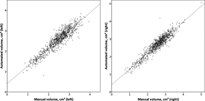
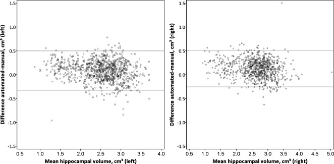
Key findings: The automated segmentation algorithm provided robust delineation of the hippocampi on 3T scans with no more variability than that seen between different human raters (Dice coefficients: interrater 0.832, manual vs. automated 0.847). In addition, the algorithm provided excellent results with the 1.5T scans (Dice coefficient 0.827), and automated segmentation remained accurate even in small sclerotic hippocampi. There was a strong correlation between manual and automated hippocampal volumes (Pearson correlation coefficient 0.929 on the left and 0.941 on the right in 3T scans).
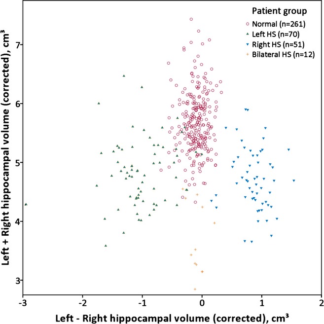
Significance: We demonstrate reliable identification of hippocampal atrophy in patients with hippocampal sclerosis, which is crucial for clinical management of epilepsy, particularly if surgical treatment is being contemplated. We provide a free online Web-based service to enable hippocampal volumetry to be available globally, with consequent greatly improved evaluation of those with epilepsy.
Keywords: Epilepsy surgery; Hippocampal sclerosis; Hippocampal segmentation; Magnetic resonance imaging.
Wiley Periodicals, Inc. © 2013 The Authors. Epilepsia published by Wiley Periodicals, Inc. on behalf of International League Against Epilepsy.
Figures
References
-
- Apostolova LG, Dinov ID, Dutton RA, Hayashi KM, Toga AW, Cummings JL, Thompson PM. 3D comparison of hippocampal atrophy in amnestic mild cognitive impairment and Alzheimer's disease. Brain. 2006;129:2867–2873. - PubMed
-
- Bernasconi N, Bernasconi A, Caramanos Z, Antel SB, Andermann F, Arnold DL. Mesial temporal damage in temporal lobe epilepsy: a volumetric MRI study of the hippocampus, amygdala and parahippocampal region. Brain. 2003;126:462–469. - PubMed
-
- Bernasconi N, Kinay D, Andermann F, Antel S, Bernasconi A. Analysis of shape and positioning of the hippocampal formation: an MRI study in patients with partial epilepsy and healthy controls. Brain. 2005;128:2442–2452. - PubMed
-
- Bremner JD, Narayan M, Anderson ER, Staib LH, Miller HL, Centerney DS. Hippocampal volume reduction in major depression. Am J Psychiatry. 2000;157:115–118. - PubMed
Publication types
MeSH terms
Grants and funding
LinkOut - more resources
Full Text Sources
Other Literature Sources
Medical
