. 2014 Feb;15(2):157-78.
doi: 10.1111/tra.12134. Epub 2013 Nov 18.

Rab6 dependent post-Golgi trafficking of HSV1 envelope proteins to sites of virus envelopment

Affiliations

Rab6 dependent post-Golgi trafficking of HSV1 envelope proteins to sites of virus envelopment

Helen L Johns et al. Traffic. 2014 Feb.

Abstract

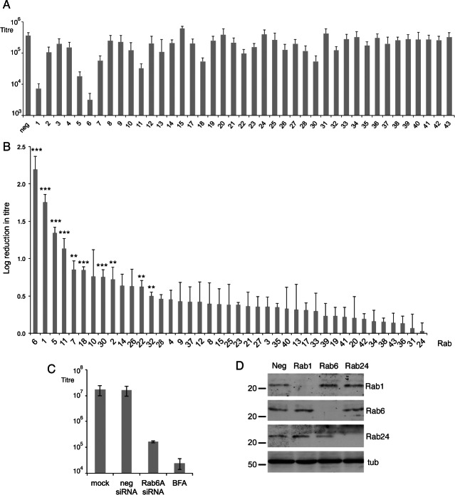
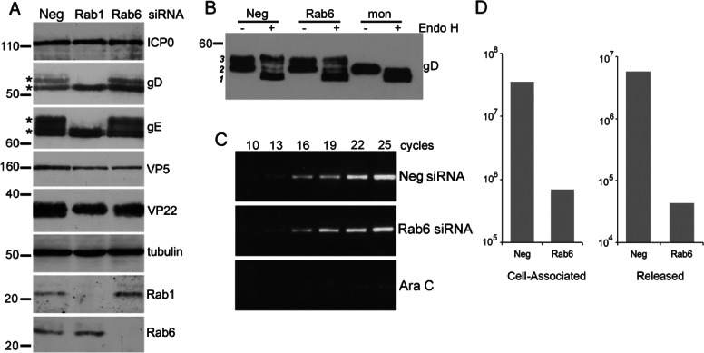
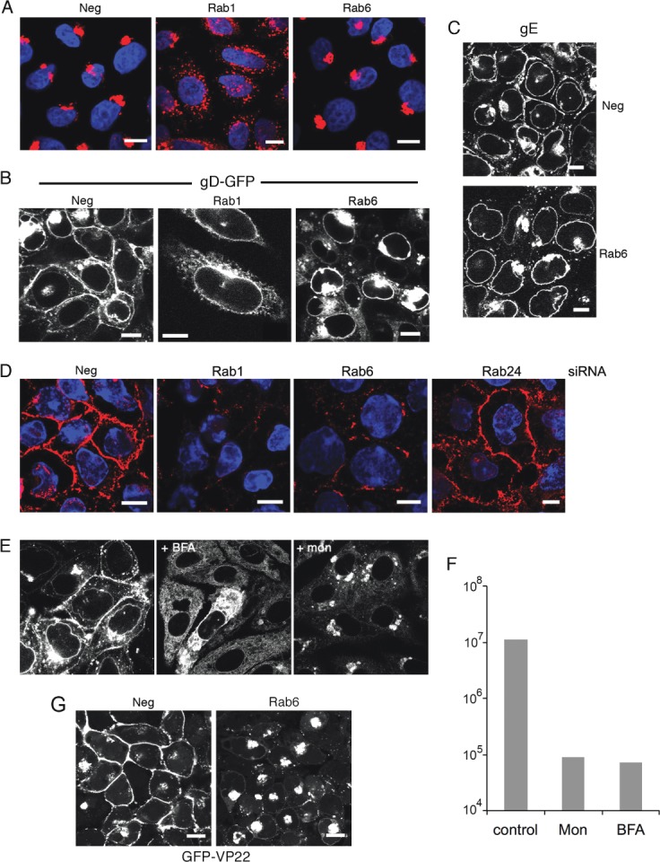
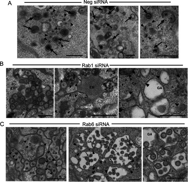
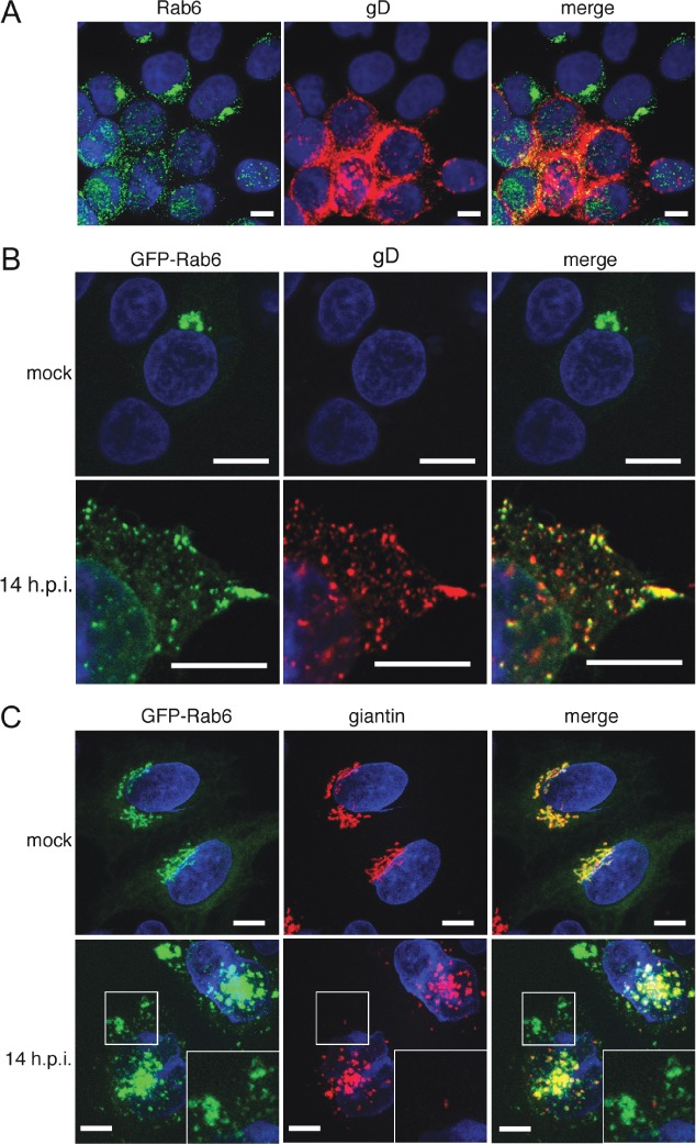
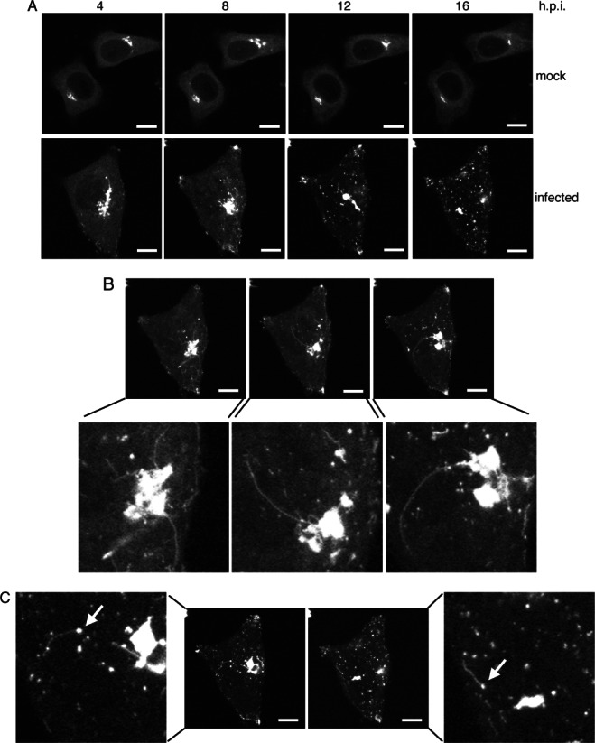
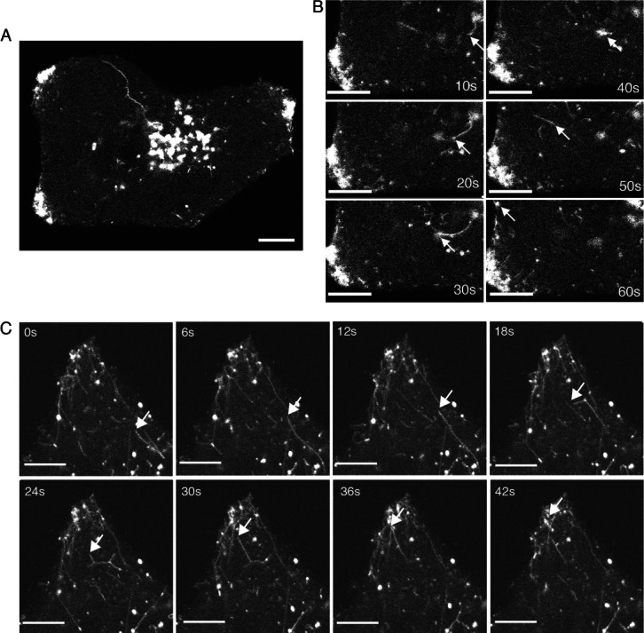
Herpes simplex virus 1 (HSV1) is an enveloped virus that uses undefined transport carriers for trafficking of its glycoproteins to envelopment sites. Screening of an siRNA library against 60 Rab GTPases revealed Rab6 as the principal Rab involved in HSV1 infection, with its depletion preventing Golgi-to-plasma membrane transport of HSV1 glycoproteins in a pathway used by several integral membrane proteins but not the luminal secreted protein Gaussia luciferase. Knockdown of Rab6 reduced virus yield to 1% and inhibited capsid envelopment, revealing glycoprotein exocytosis as a prerequisite for morphogenesis. Rab6-dependent virus production did not require the effectors myosin-II, bicaudal-D, dynactin-1 or rabkinesin-6, but was facilitated by ERC1, a factor involved in linking microtubules to the cell cortex. Tubulation and exocytosis of Rab6-positive, glycoprotein-containing membranes from the Golgi was substantially augmented by infection, resulting in enhanced and targeted delivery to cell tips. This reveals HSV1 morphogenesis as one of the first biological processes shown to be dependent on the exocytic activity of Rab6.

Keywords: Gaussia luciferase; Rab6; Rab6 effector; VSV-G; herpes simplex virus; post-Golgi carrier; secretion.


Figures

Figure 1
Figure 1
Requirement for human Rab GTPases in HSV1 infection. A) siRNAs from a library against 60 human Rabs were transfected into HeLa cells, alongside a control negative siRNA, pooling siRNAs to different isoforms of the same Rab. Two days later, the transfected cells were infected with HSV1 at a multiplicity of 2, and extracellular released virus harvested at 16 h and titrated on Vero cells. Error bars indicate standard error from three independent experiments. B) The graph from (A) is presented as the relative log drop in virus yield, in order from the largest to the smallest. Error bars indicate standard error from three independent experiments. Statistical analysis was carried out using the Student's t-test. ***p < 0.001; **p < 0.01. C) HeLa cells were transfected with negative or Rab6A siRNAs or left untransfected, and infected as above. Five hour after infection BFA (1 µg/mL) was added to one untransfected sample, and released virus harvested from all samples at 18 h. Error bars indicate standard error from three independent experiments. D) HeLa cells were transfected with the negative siRNA or pooled siRNAs to Rab1A and1B, Rab6A, 6B and 6C or Rab24 and analysed 2 days later by western blotting for these Rabs. Alpha tubulin (tub) was used as the loading control. Molecular weight markers are shown in kDa.
Figure 2
Figure 2
Rab6 depletion inhibits virus production at late stages of infection. A) HeLa cells transfected with neg, Rab1 or Rab6 siRNAs were infected with HSV1 2 days later and harvested 16 h after infection for western blotting with antibodies as indicated. Mature glycosylated forms of gD and gE are marked by asterisk. B) The status of glycoprotein glycosylation in control (neg), Rab6 depleted or monensin treated cells was determined by Endo H treatment of infected cell lysates harvested at 16 h, and analysed by western blotting for gD. Three isoforms of gD are denoted by 1, 2 & 3. C) The level of DNA replication in control (neg), Rab6 depleted or Ara C treated cells was analysed by semi-quantitative PCR using a primer pair specific for the UL47 gene. PCR cycle numbers are denoted. D) Hela cells transfected with neg or Rab6 siRNAs were infected 2 days later with HSV1, and cell-associated and released virus titrated on Vero cells. Molecular weight markers are shown in kDa.
Figure 3
Figure 3
Rab6 depletion inhibits HSV1 glycoprotein localization to the plasma membrane in infected cells. A) HeLa cells grown on coverslips were transfected with neg, Rab1 or Rab6 siRNAs, fixed 2 days later and stained for giantin (red), and nuclei stained wih DAPI (blue). B) HeLa cells on coverslips transfected with neg, Rab1 or Rab6 siRNAs were infected 2 days later with HSV1 expressing gD-GFP and fixed 16 h later. C) Neg or Rab6 siRNA transfected HeLa cells were infected after 2 days with Wt HSV1 and stained with a monoclonal antibody against gE. D) HeLa cells on coverslips were transfected with neg, Rab1, Rab6 or Rab24 siRNAs and infected 2 days later with Wt HSV1. Sixteen hours later the cells were incubated on ice for 30 min with gD monoclonal antibody to detect cell surface protein, washed and fixed prior to staining with secondary antibody (red). Nuclei were stained with DAPI (blue). E) Cells infected with HSV1 expressing gD-GFP at a multiplicity of 2 were treated 5 h later with BFA, monensin or left untreated. Cells were fixed at 12 h. F) Total virus yield from infections carried out as for (E), and harvested 18 h after infection. G) HeLa cells on coverslips were transfected with neg or Rab6 siRNA, infected 2 days later with HSV1 expressing the tegument protein VP22 as a GFP fusion protein (GFP-VP22), and images acquired after 16 h. Scale bar = 10 µm.
Figure 4
Figure 4
Rab6 depletion inhibits HSV1 capsid envelopment. Hela cells transfected with (A) neg, (B) Rab1 or (C) Rab6 siRNAs were infected 2 days later with HSV1, fixed at 16 h and processed for transmission electron microscopy. ER, endoplasmic reticulum; GA, Golgi apparatus; Ca, capsid. Arrowheads indicate naked capsids. Thick arrows indicate capsids associated with wrapping membranes. Thin arrows indicate continuum with the cytoplasm. Scale bar = 500 nm.
Figure 5
Figure 5
Rab6 is redistributed and colocalizes with HSV1 glycoproteins at the cell periphery. A) HeLa cells infected with HSV1 at a multiplicity of 0.5 were fixed 16 h later and stained for endogenous Rab6 (green) and gD (red). Nuclei were stained with DAPI (blue). B and C) HeLa cells transfected with plasmid expressing GFP-Rab6 (green) were mock or HSV1 infected, fixed 14 h later and stained with antibodies for the glycoprotein gD (B) or the Golgi marker giantin (C) (red). Nuclei were stained with DAPI (blue). Scale bar = 10 µm.
Figure 6
Figure 6
Rab6 depletion inhibits trafficking of gD, gE and tsVSV-G, but not Gaussia luciferase. A) HeLa cells on coverslips were transfected with neg, Rab1 or Rab6 siRNAs and 2 days later transfected with plasmids expressing gD or gE. After 16 h the cells were processed for cell surface staining with the appropriate antibody, incubated with secondary antibody (red), and stained with DAPI (blue). B) HeLa cells on coverslips were transfected with neg, Rab1 or Rab6 siRNAs and 2 days later transfected with plasmid expressing GFP-tsVSV-G protein (green). The cells were incubated overnight at the non-permissive temperature of 39°C (pre-shift), then incubated at the permissive temperature of 32°C for 4 h (post-shift) before fixation and staining with DAPI (blue). C) HeLa cells treated as indicated were transfected with a plasmid expressing Gaussia luciferase and incubated for 16 h. Media was changed and 1 h later the level of secreted Gaussia luciferase was measured, with the amount secreted from cells transfected with the neg siRNA taken as 1. As a positive control BFA was added to cells at a concentration of 5 µg/mL before assay. Un, untransfected cells. Error bars indicate standard error from three experiments. Scale bar = 10 µm.
Figure 7
Figure 7
The Rab6 fission effector non-muscle myosin II is not required for HSV1 morphogenesis. A) HeLa cells infected with HSV1 at a multiplicity of 2 were treated with blebbistatin at 5 h or left untreated and harvested for western blotting at 16 h. B) HeLa or HFFF cells infected with HSV1 at a multiplicity of 2 were treated with blebbistatin at 5 h or left untreated and harvested for extracellular virus yield at 18 h. C and D) HeLa cells were transfected with neg, myosin IIA (IIA) or myosin IIB (IIB) siRNAs, infected 2 day later with HSV1 at a multiplicity of 2, and harvested for western blotting (C) or extracellular virus (D). E) HeLa cells on coverslips were transfected with neg or myosin IIB (NMIIB) siRNAs and infected 2 days later with Wt HSV1. Eight and sixteen hours later the cells were processed for gD detection at the cell surface, and stained with secondary antibody (red). Nuclei were stained with DAPI (blue). Scale bar = 10 µm. Error bars indicate standard error from three experiments. Molecular weight markers are shown in kDa.
Figure 8
Figure 8
The Rab6 effector ERC1 facilitates HSV1 glycoprotein trafficking and morphogenesis. A–C) HeLa cells were transfected with neg siRNA or siRNA to bicaudal D1 (BicD1), bicaudal D2 (BicD2), conventional kinesin heavy chain (KIF5B), dynactin 1 (Dyn), ERC1 or rabkinesin 6 (KIF20A) and 2 days later were either harvested for western blotting with antibodies specific to each effector (A) or infected with HSV1 at a multiplicity of 2, and harvested at 16 h for western blotting for the virus proteins ICP0 and gD (B), or extracellular virus (C). Error bars indicate standard error from three experiments. D) HeLa cells on coverslips were transfected with neg or ERC1 siRNAs and infected 2 days later with HSV1. Eight or sixteen hours later the cells were processed for detection of cell surface gD, and stained with secondary antibody (red). Nuclei were stained with DAPI (blue), and images were acquired with the same acquisition settings. Scale bar = 10 µm. Molecular weight markers are shown in kDa.
Figure 9
Figure 9
Rab6 trafficking to the cell surface is activated in HSV1 infected cells. HeLa cells were transfected with plasmid expressing GFP-Rab6 and left uninfected (mock) or infected 4 h later with HSV1 (infected). After a further 4 h, time-lapse analysis was carried out with images acquired every 5 min for 16 h. A) Images acquired at 4 hourly intervals through the course of the time-lapse showing the progressive dispersal of GFP-Rab6 through infection. B and C) Representative images showing GFP-Rab6 positive tubules emanating from the area of the Golgi/TGN (B) or GFP-Rab6 positive domains in cytoplasm (C). Arrows indicate peripheral sites from where tubules extend. Scale bar = 10 µm. See also Video S1.
Figure 10
Figure 10
Enhanced exocytosis of Rab6 tubules in HSV1 infected cells. A and B) HeLa cells were transfected with plasmid expressing GFP-Rab6 and infected 12 h later with HSV1 at a multiplicity of 2. After 8 h (A and B) or 16 h (C), images were acquired at (B) 10 seconds or (C) 1 seconds intervals. Scale bar = 10 µm. See also Videos S2–S4.

Similar articles

Cited by

References

    1. Stenmark H. Rab GTPases as coordinators of vesicle traffic. Nat Rev Mol Cell Biol. 2009;10:513–525. - PubMed
    1. Hutagalung AH, Novick PJ. Role of Rab GTPases in membrane traffic and cell physiology. Physiol Rev. 2011;91:119–149. - PMC - PubMed
    1. Utley TJ, Ducharme NA, Varthakavi V, Shepherd BE, Santangelo PJ, Lindquist ME, Goldenring JR, Crowe JE., Jr Respiratory syncytial virus uses a Vps4-independent budding mechanism controlled by Rab11-FIP2. Proc Natl Acad Sci USA. 2008;105:10209–10214. - PMC - PubMed
    1. Amorim MJ, Bruce EA, Read EK, Foeglein A, Mahen R, Stuart AD, Digard P. A Rab11- and microtubule-dependent mechanism for cytoplasmic transport of influenza A virus viral RNA. J Virol. 2011;85:4143–4156. - PMC - PubMed
    1. Bruce EA, Digard P, Stuart AD. The Rab11 pathway is required for influenza A virus budding and filament formation. J Virol. 2010;84:5848–5859. - PMC - PubMed

Publication types

MeSH terms