. 2013 Oct 23;14(1):113.
doi: 10.1186/1465-9921-14-113.

Pharmacological inhibition of GSK-3 in a guinea pig model of LPS-induced pulmonary inflammation: I. Effects on lung remodeling and pathology

Affiliations

Pharmacological inhibition of GSK-3 in a guinea pig model of LPS-induced pulmonary inflammation: I. Effects on lung remodeling and pathology

Hoeke A Baarsma et al. Respir Res. .

Abstract

Background: Glycogen synthase kinase-3 (GSK-3) is a constitutively active kinase that regulates multiple signalling proteins and transcription factors involved in a myriad of cellular processes. The kinase acts as a negative regulator in β-catenin signalling and is critically involved in the smad pathway. Activation of both pathways may contribute to pulmonary features of chronic obstructive pulmonary disease (COPD).

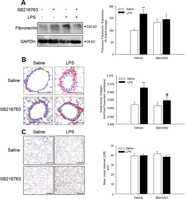
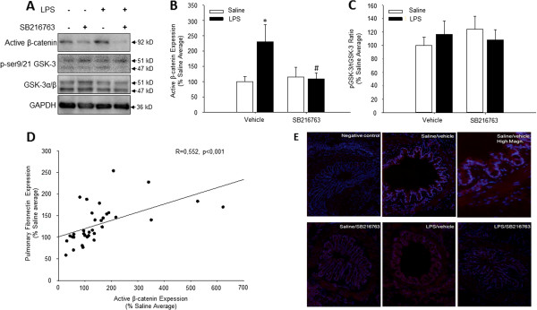
Methods: In the present study, we investigated the effect of the selective GSK-3 inhibitor SB216763 on pulmonary pathology in a guinea pig model of lipopolysaccharide (LPS)-induced COPD. Guinea pigs were instilled intranasally with LPS or saline twice weekly for 12 weeks and pre-treated with either intranasally instilled SB216763 or corresponding vehicle 30 min prior to each LPS/saline challenge.

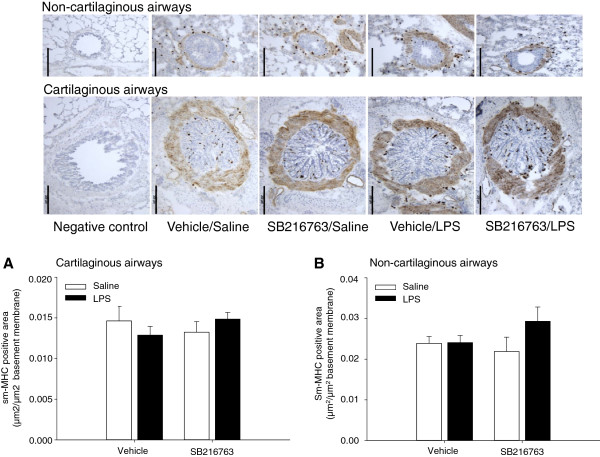
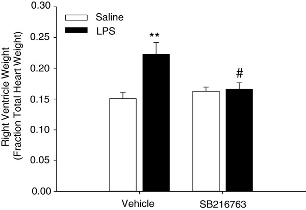
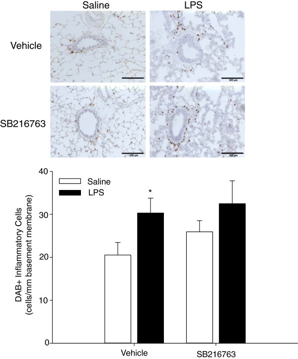
Results: Repeated LPS exposures activated β-catenin signalling, primarily in the airway epithelium and submucosa. LPS also induced pulmonary inflammation and tissue remodelling as indicated by inflammatory cell influx, increased pulmonary fibronectin expression and enhanced small airway collagen content. Inhibition of GSK-3 by SB216763 did not affect LPS-induced inflammatory cell influx, but prevented the small airway remodelling and, unexpectedly, inhibited the activation of β-catenin in vivo. LPS or SB216763 treatment had no effect on the airway smooth muscle content and alveolar airspace size. However, GSK-3 inhibition prevented LPS-induced right ventricle hypertrophy.

Conclusions: Our findings indicate that GSK-3 inhibition prevents LPS-induced pulmonary pathology in guinea pigs, and that locally reduced LPS-induced β-catenin activation appears in part to underlie this effect.


Figures

Figure 1
Figure 1
Effect of repeated intranasal LPS challenge and treatment with the selective GSK-3 inhibitor SB216763 on extracellular matrix deposition in the lung. (A) Expression of fibronectin was evaluated in whole lung homogenates 24 hours after the last challenge by immunoblotting using specific antibodies. Equal protein loading was verified by the analysis of GAPDH. Effects of repeated LPS challenge and SB216763 treatment on fibronectin expression were quantified by densitometry, representing mean ± s.e.m. of 9 animals per group. (B) Histological staining of the extracellular matrix protein collagen using Sirius Red. The non-cartilaginous airways were digitally photographed (100-200 × magnification) and analysed by using ImageJ software. Effects of repeated LPS challenge and SB216763 treatment on airway collagen expression were quantified, representing mean ± s.e.m. of 9 animals per group. (C) The mean linear intercept (LMI), a measure for alveolar airspace size, was determined by staining the tissue-sections with haematoxylin and eosin. Data represent means ± s.e.m. of 9 animals per group. **p < 0.01 compared to control group and #p < 0.05 compared to LPS treated animals. Scale bar = 200 μm.
Figure 2
Figure 2
Repeated LPS instillation and pharmacological inhibition of GSK-3 by SB216763 do not affect airway smooth muscle content. Immunohistological analysis of sm-MHC positive area in (A) large (cartilaginous) and (B) small (non-cartilaginous) airways. Effects of repeated LPS challenge and SB216763 treatment on airway smooth muscle sm-MHC expression were quantified. Data represent means ± s.e.m. of 9 animals per group. Scale bar = 200 μm.
Figure 3
Figure 3
Right ventricle hypertrophy after repeated intranasal LPS instillation is attenuated by GSK-3 inhibition. Effect of repeated LPS instillation and GSK-3 inhibition by SB216763 on right ventricle hypertrophy. Effects of repeated LPS challenge and SB216763 treatment on size of right ventricle were quantified as right ventricle weight over total heart weight, representing mean ± s.e.m. of 9 animals per group. **p < 0.01 compared to control group and #p < 0.05 compared to LPS treated animals.
Figure 4
Figure 4
GSK-3 inhibition does not inhibit LPS-induced pulmonary inflammation. Effect of repeated LPS instillation and GSK-3 inhibition by SB216763 on inflammatory cell infiltration in the airways. Cells within 50 μm of the basement membrane were quantified and expressed relative to basement membrane length, representing mean ± s.e.m. of 9 animals per group. *p < 0.05 compared to control group. Scale bar = 200 μm.
Figure 5
Figure 5
Activation of β-catenin in response to repeated intranasal LPS challenge is prevented by treatment with the selective GSK-3 inhibitor SB216763. (A) Expression of active β-catenin, phosphorylated GSK-3 (ser9/21 GSK-3) and total GSK-3 was evaluated in whole lung homogenates 24 hours after the last challenge by immunoblotting using specific antibodies. Equal protein loading was verified by the analysis of GAPDH. (B,C) Responses of repeated LPS challenge and SB216763 treatment on active β-catenin expression (B) and GSK-3 phosphorylation (C) were quantified by densitometry, representing mean ± s.e.m. of 9 animals per group. (D) Correlation between pulmonary expression of fibronectin (data from Figure 1) and active β-catenin in all guinea pigs. R = 0.552; p < 0.001. (E) Immunofluorescence analysis of active β-catenin (red) in large airways counterstained with Hoechst 3342 to stain nuclei (blue). *p < 0.05 compared to control group and #p < 0.05 compared to LPS treated animals.

Similar articles

Cited by

References

    1. Woodgett JR. cDNA cloning and properties of glycogen synthase kinase-3. Meth Enzymol. 1991;14:564–577. - PubMed
    1. Woodgett JR. Molecular cloning and expression of glycogen synthase kinase-3/factor A. EMBO J. 1990;14(8):2431–2438. - PMC - PubMed
    1. Rayasam GV, Tulasi VK, Sodhi R, Davis JA, Ray A. Glycogen synthase kinase 3: more than a namesake. Br J Pharmacol. 2009;14(6):885–898. doi: 10.1111/j.1476-5381.2008.00085.x. - DOI - PMC - PubMed
    1. Embi N, Rylatt DB, Cohen P. Glycogen synthase kinase-3 from rabbit skeletal muscle. Separation from cyclic-AMP-dependent protein kinase and phosphorylase kinase. Eur J Biochem. 1980;14(2):519–527. - PubMed
    1. Frame S, Cohen P. GSK3 takes centre stage more than 20 years after its discovery. Biochem J. 2001;14:1–16. doi: 10.1042/0264-6021:3590001. - DOI - PMC - PubMed

Publication types

MeSH terms