. 2013 Nov 6;135(44):16517-25.
doi: 10.1021/ja407451c. Epub 2013 Oct 23.

Translocation of cationic amphipathic peptides across the membranes of pure phospholipid giant vesicles

Affiliations

Translocation of cationic amphipathic peptides across the membranes of pure phospholipid giant vesicles

Sterling A Wheaten et al. J Am Chem Soc. .

Abstract

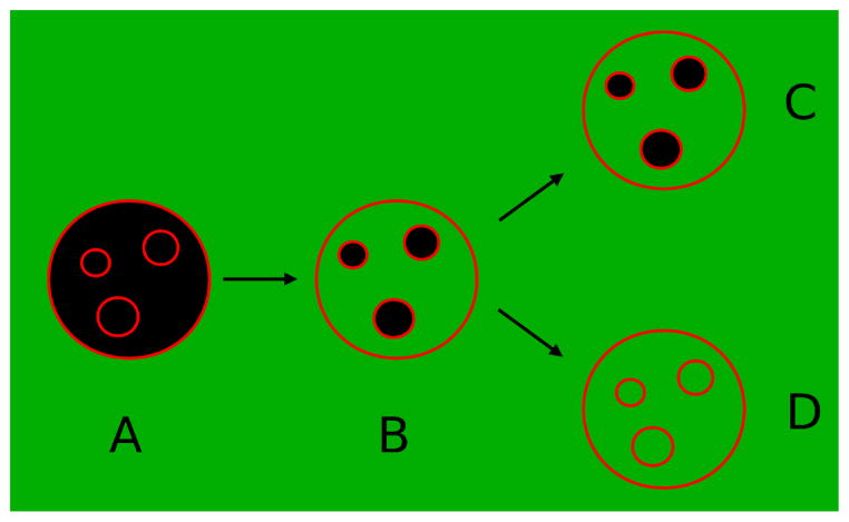
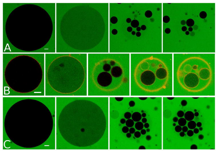
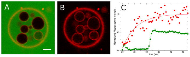
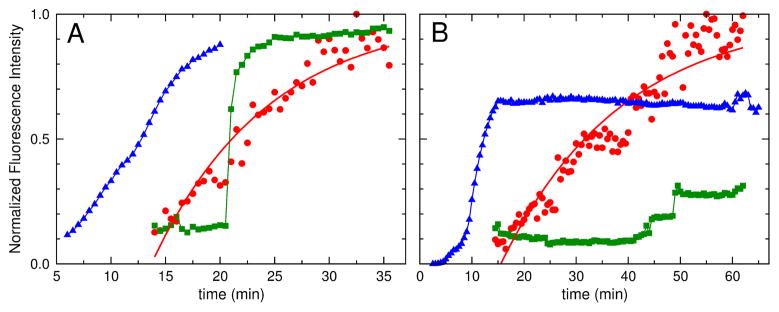
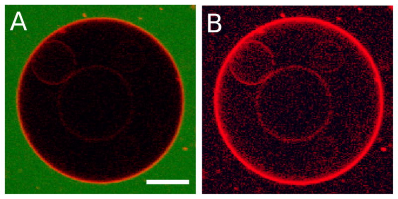
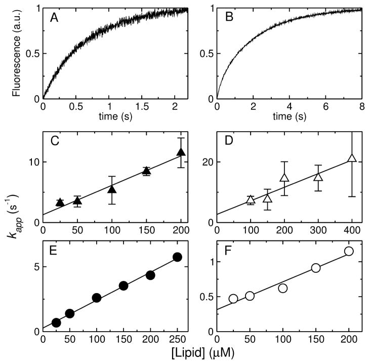
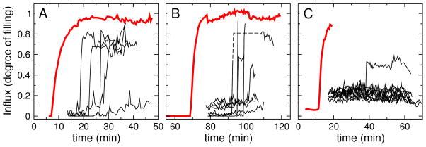
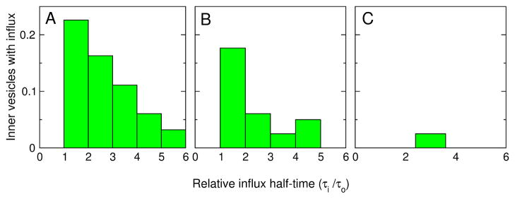
The ability of amphipathic polypeptides with substantial net positive charges to translocate across lipid membranes is a fundamental problem in physical biochemistry. These peptides should not passively cross the bilayer nonpolar region, but they do. Here we present a method to measure peptide translocation and test it on three representative membrane-active peptides. In samples of giant unilamellar vesicles (GUVs) prepared by electroformation, some GUVs enclose inner vesicles. When these GUVs are added to a peptide solution containing a membrane-impermeant fluorescent dye (carboxyfluorescein), the peptide permeabilizes the outer membrane, and dye enters the outer GUV, which then exhibits green fluorescence. The inner vesicles remain dark if the peptide does not cross the outer membrane. However, if the peptide translocates, it permeabilizes the inner vesicles as well, which then show fluorescence. We also measure translocation, simultaneously on the same GUV, by the appearance of fluorescently labeled peptides on the inner vesicle membranes. All three peptides examined are able to translocate, but to different extents. Peptides with smaller Gibbs energies of insertion into the membrane translocate more easily. Further, translocation and influx occur broadly over the same period, but with very different kinetics. Translocation across the outer membrane follows approximately an exponential rise, with a characteristic time of 10 min. Influx occurs more abruptly. In the outer vesicle, influx happens before most of the translocation. However, some peptides cross the membrane before any influx is observed. In the inner vesicles, influx occurs abruptly sometime during peptide translocation across the membrane of the outer vesicle.


Conflict of interest statement

Notes

The authors declare no competing financial interests.

Figures

Figure 1
Figure 1
The concept of the experiment. GUVs prepared with inner vesicles are added to a solution containing peptide and a water-soluble fluorophore (green). The membrane of the vesicles is shown in red. Initially (A), the interior of the vesicles has no fluorescence (black). In (B) the peptide induced flux into the outer vesicle. If the peptide does not translocate across the outer membrane, the inner vesicles remain dark (C). Appearance of fluorescence inside inner vesicles indicates that the peptide translocated across the membrane of the outer vesicle (D).
Figure 2
Figure 2
Sequences of CF (green fluorescence) influx into POPC GUVs as a function of time upon addition to a solution of (A and B) Rh-TP10W or (C) Rh-DL-1a. In (B) the amplification of the rhodamine channel was increased to show the rhodamine labeled peptide on the membrane of the inner vesicles (last images of the series). The times of each frame, from the moment of addition of the GUVs to the peptide are (A) 11.5, 21.5, 25, and 29 min; (B) 8.5, 14, 33, 60, and 75 min; (C) 14, 19, 27, and 28–69 min. Peptide concentration = 0.75 μM, lipid concentration ~ 50 μM. Scale bar, 10 μm.
Figure 3
Figure 3
Large GUV with inner vesicles 44 min after addition of the peptide Rh-TP10W. (A) CF and Rh channels. (B) Rh channel only. Scale bar, 10 μm. (C) Fluorescence of the inner vesicle at the bottom right in A and B, as a function of time. Red points, Rh channel, showing Rh-TP10W on the inner vesicle membrane. Green points, CF channel, showing flux into the inner vesicle (intensity relative to the outer membrane). The red line is 1 − exp[−(tto)/τR] where τR = 5.8 min and to ≈ 20 min (beginning of recording). The green line is only to guide the eye.
Figure 4
Figure 4
Flux and translocation as a function of time in two different vesicles (A,B) upon addition to Rh-TP10W. Data and fits as in Fig. 3. Red, translocation across the outer vesicle membrane. Green, dye flux into the inner vesicle. Blue, dye flux into the outer GUV. Fits (red lines) yield (A) τR = 6.6 min and (B) τR = 23 min. Vesicle (B) moved out of the focal plane at the end of the period shown.
Figure 5
Figure 5
Vesicle at 74 min from beginning of experiment showing inner vesicles with Rh-TP10W on their membranes (A) CF and Rh channels. No CF influx into the outer vesicle was observed. (B) Rh channel, enhanced, showing inner vesicles with Rh-TP10W. Scale bar, 20 μm.
Figure 6
Figure 6
Examples of binding kinetics curves for (A) Rh-TP10W and (B) Rh-DL-1a, with 1 μM peptide and 50 μM lipid concentrations, and plots of kapp as a function of lipid concentration for (C) Ac-TP10W, (D) Ac-DL-1a, (E) Rh-TP10W, and (F) Rh-DL-1a. In (E) and (F) the error bars are inside the points.
Figure 7
Figure 7
Time traces of CF flux into inner GUVs (black) compared with the influx into the outer GUV (red). (A) Rh-TP10W, (B) Rh-DL-1, and (C) CE-2. The fluorescence intensity inside the vesicles is measured relative to an area outside the vesicles. The dashed line in (B) traces a vesicle whose track was temporarily lost, as it drifted out of focus.
Figure 8
Figure 8
Density distributions of influx half-times of the inner vesicles for (A) Rh-TP10W, (B) Rh-DL-1a, and (C) CE-2. The times of the midpoints in the influx curves (Figure 7) of the inner vesicles (τi) are expressed relative to their outer vesicle (τo) in each sample, and all inner vesicles are pooled for each peptide.

References

    1. Langel U. Handbook of Cell Penetrating Peptides. 2. CRC Press; Boca Raton, FL: 2006.
    1. Derossi D, Calvet S, Trembleau A, Brunissen A, Chassaing G, Prochiantz A. J Biol Chem. 1996;271:18188–18193. - PubMed
    1. Green M, Weston PM. Cell. 1988;55:1179–1188. - PubMed
    1. Frankel AD, Pabo CO. Cell. 1988;55:1189–1193. - PubMed
    1. Yandek LE, Pokorny A, Floren A, Knoelke K, Langel U, Almeida PFF. Biophys J. 2007;92:2434–2444. - PMC - PubMed

Publication types

LinkOut - more resources