. 2014 Mar 20;20(9):1365-81.
doi: 10.1089/ars.2013.5326. Epub 2013 Oct 23.

Mass spectrometry-based quantitative proteomics for dissecting multiplexed redox cysteine modifications in nitric oxide-protected cardiomyocyte under hypoxia

Affiliations

Mass spectrometry-based quantitative proteomics for dissecting multiplexed redox cysteine modifications in nitric oxide-protected cardiomyocyte under hypoxia

Kuan-Ting Pan et al. Antioxid Redox Signal. .

Abstract

Aims: Distinctive states of redox-dependent cysteine (Cys) modifications are known to regulate signaling homeostasis under various pathophysiological conditions, including myocardial injury or protection in response to ischemic stress. Recent evidence further implicates a dynamic interplay among these modified forms following changes in cellular redox environment. However, a precise delineation of multiplexed Cys modifications in a cellular context remains technically challenging. To this end, we have now developed a mass spectrometry (MS)-based quantitative approach using a set of novel iodoacetyl-based Cys-reactive isobaric tags (irreversible isobaric iodoacetyl Cys-reactive tandem mass tag [iodoTMT]) endowed with unique irreversible Cys-reactivities.

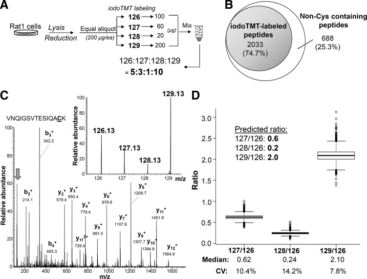
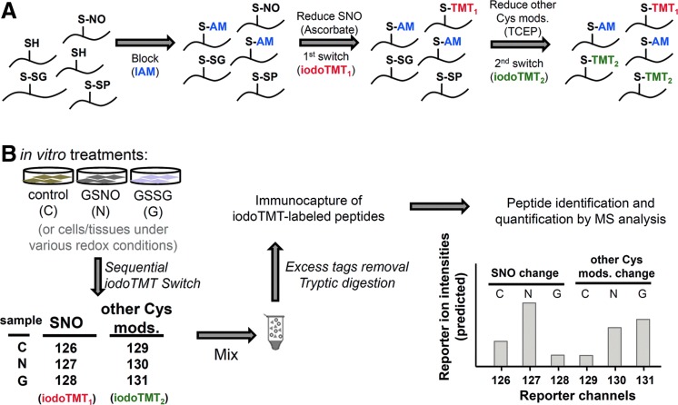
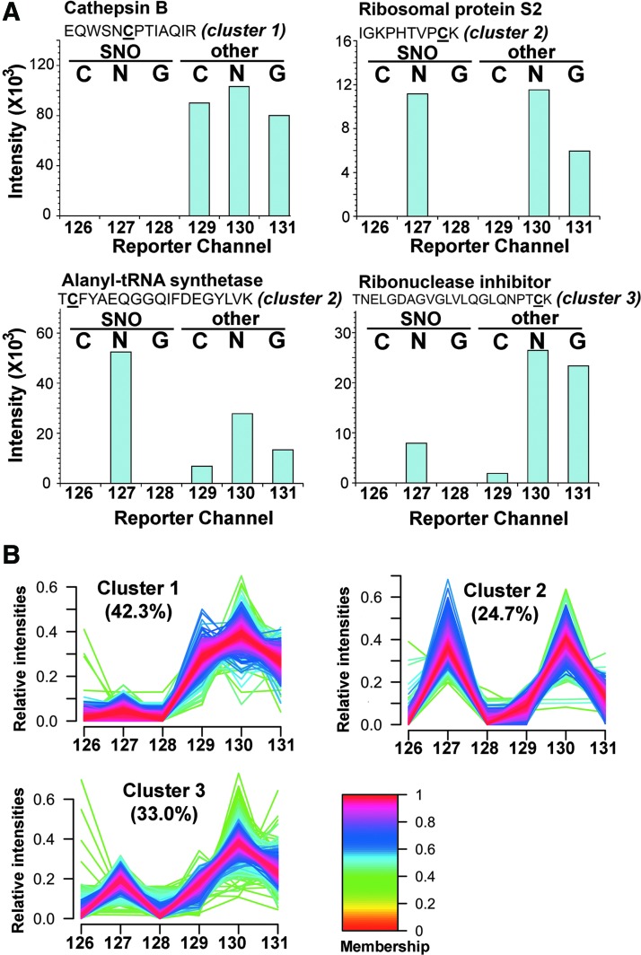
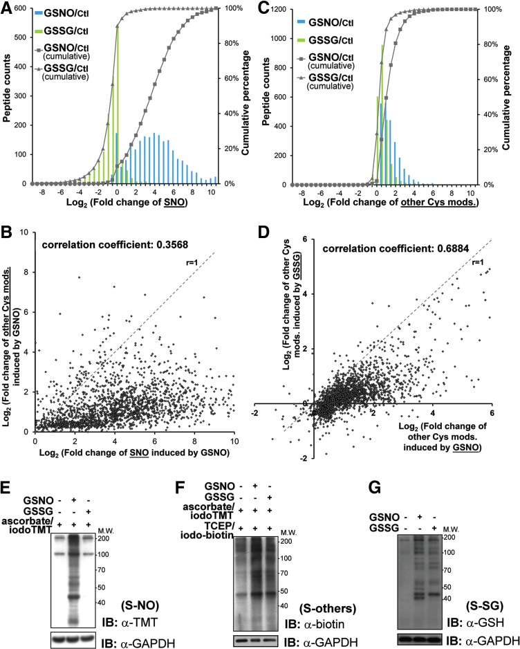
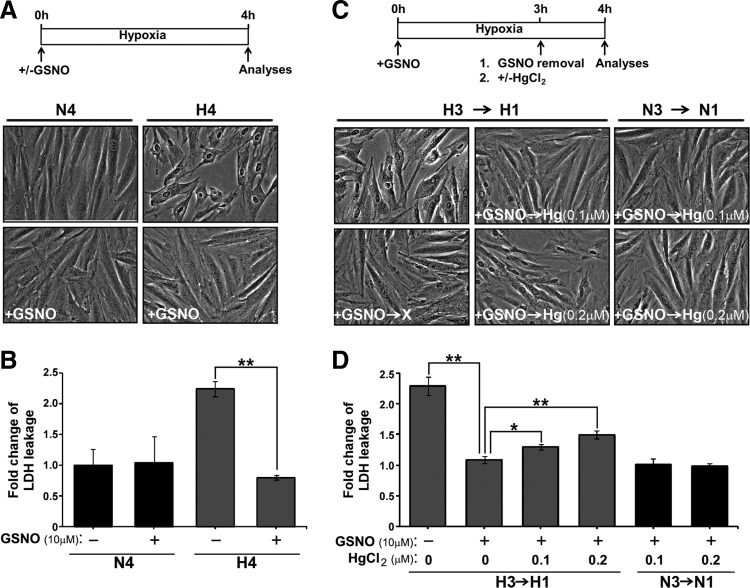
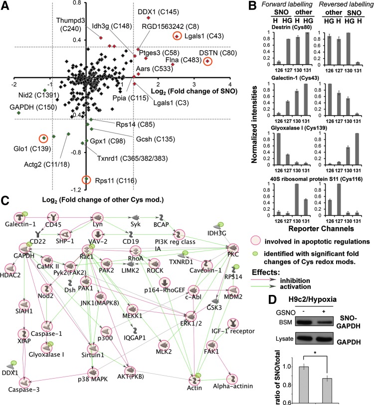
Results: We have established a sequential iodoTMT-switch procedure coupled with efficient immunoenrichment and advanced shotgun liquid chromatography-MS/MS analysis. This workflow allows us to differentially quantify the multiple redox-modified forms of a Cys site in the original cellular context. In one single analysis, we have identified over 260 Cys sites showing quantitative differences in multiplexed redox modifications from the total lysates of H9c2 cardiomyocytes experiencing hypoxia in the absence and presence of S-nitrosoglutathione (GSNO), indicative of a distinct pattern of individual susceptibility to S-nitrosylation or S-glutathionylation. Among those most significantly affected are proteins functionally implicated in hypoxic damage from which we showed that GSNO would protect.

Innovation: We demonstrate for the first time how quantitative analysis of various Cys-redox modifications occurring in biological samples can be performed precisely and simultaneously at proteomic levels.

Conclusion: We have not only developed a new approach to map global Cys-redoxomic regulation in vivo, but also provided new evidences implicating Cys-redox modifications of key molecules in NO-mediated ischemic cardioprotection.


Figures

<b>FIG. 1.</b>
FIG. 1.
Chemical structures of isobaric iodoacetyl tandem mass tag sixplex reagents (iodoTMT6). Each of the isobaric iodoTMT6 reagents has the same nominal mass and comprises a sulfhydryl-reactive iodoacetyl group, a mass normalizing spacer arm and a mass reporter. Asterisks (*) indicate the positions in each of the mass tags where 12C and 14N are replaced by 13C and 15N. Under MS/MS conditions, the isobaric mass tags are readily cleaved at the bond indicated by dash line, thus generating reporter ions with unique m/z of 126–131. iodoTMT, irreversible isobaric iodoacetyl Cys-reactive tandem mass tag; MS, mass spectrometry.
<b>FIG. 2.</b>
FIG. 2.
The accuracy and precision of iodoTMT-based multiplexed quantification. (A) Schematics of the sample preparation strategy used to evaluate the performance of iodoTMT-based quantification as mentioned in text. After tryptic digestion, the anti-TMT affinity enriched peptides were subjected to liquid chromatography (LC)-MS/MS analysis, which resulted in identification of over 2000 iodoTMT-labeled peptides representing an enrichment specificity of ∼75% (B). A representative MS/MS spectrum of an iodoTMT-labeled peptide is shown in (C), with its major sequence-informative b/y fragment ions fully annotated and the narrow mass region containing the TMT reporter ions (marked by an arrow) further expanded in the inset. Deviation of the experimentally derived reporter ion ratio values from the predicted ratios, namely 0.6, 0.2, and 2.0, for m/z 127, 128, and 129 to 126, respectively, for all 1669 quantifiable iodoTMT-labeled peptides as shown by Boxplot in (D), was taken as a measure of the quantification accuracy. CV, coefficient of variation.
<b>FIG. 3.</b>
FIG. 3.
Experimental workflow of the sequential iodoTMT switch strategy for simultaneous monitoring of multiple Cys-modifications and quantitative determination of their respective ratios. (A) The principle requires that samples to be assayed be first treated with IAM to block all SH. Subsequently, S-nitrosylated Cys residues (SNO) are specifically reduced by ascorbate followed by iodoTMT1 labeling. Both S-SG and S-SP are then reduced by TCEP prior to iodoTMT2 alkylation. Note that the second switch will also convert any other reversible modifications that may be present to iodoTMT2. Since up to sixplexed quantification can be effected and the sequential switch strategy utilizes two channels per sample, up to three cell states can be monitored simultaneously. In the example illustrated in (B), the same cells can either be treated with GSNO (N) or GSSG (G), or non-treated (control, C). Following the sequential iodoTMT switch with various channels of iodoTMT tags as indicated, all samples were mixed, digested, and enriched for iodoTMT-labeled peptides prior to LC-MS/MS analysis. Relative abundance of the reporter ions detected for each positively identified peptide can then be used to calculate fold changes of SNO versus other reversible Cys modifications upon treatments with either GSNO or GSSG, relative to control. Cys, cysteine; GSNO, S-nitrosoglutathione; GSSG, glutathione disulfide; IAM, iodoacetamide; SH, free thiol; SNO, S-nitrosylation; S-SG, S-glutathionylation; S-SP, protein disulfides; TCEP, tris(2-carboxyethyl) phosphine. To see this illustration in color, the reader is referred to the web version of this article at www.liebertpub.com/ars
<b>FIG. 4.</b>
FIG. 4.
Clustering of the identified Cys residues based on the relative intensities of the reporter ions. Four representative intensity patterns of the reporter ion channels were shown in (A). Notably, the Cys in cathepsin B is known to be involved in disulfide bridge and therefore only “other” form and no SNO was detected. Good specificity of ascorbate reduction was shown by detecting SNO only in channel 127. The varying amount of SNO and other Cys-modification induced by GSNO on each specific Cys site is represented by the relative intensity ratio of reporter ions 127 to 130. The other three examples shown represent sites where approximately equal amounts (ribosomal protein S2), more SNO (alanyl-tRNA synthetase) and less SNO (ribonuclease inhibitor) than other Cys modifications were induced by added GSNO. All identified Cys residues were classified by the patterns of the relative intensities of reporter ions into three clusters using GproX (B). Percentage of the involved Cys residues in each cluster was shown as indicated with the color-coded intensities of membership shown at right.
<b>FIG. 5.</b>
FIG. 5.
Relative amount of induced SNO and other Cys-modification upon in vitro treatment of H9c2 cell lysates with GSNO or GSSG. Global analysis by the sequential iodoTMT switch method revealed that GSNO but not GSSG treatment would induce significant SNO formation as shown by the histograms of fold changes of SNO induced by different treatments (A). The solid lines with closed square or triangle labels represent the cumulative percentage of induced SNO by GSNO or GSSG treatments, respectively. This is further supported by anti-TMT immunoblotting in parallel experiments involving only the first switching step (inset, gel pattern). On average, GSNO was shown to induce more SNO formation than other Cys-modifications on most Cys sites, which led to the observed scatter plot pattern (B). Dash line in the scatter plot (slope=1) represented equal fold change of SNO and S-SG of particular Cys residues under GSNO treatment. Correlation coefficient of induced fold changes of SNO versus other Cys modifications was shown as indicated. Moreover, GSNO induced an overall higher amount of other Cys modification than GSSG did (C), as revealed by the corresponding scatter plot pattern (D). All measured fold changes were relative to untreated control. The changes of SNO and other Cys modifications caused by in vitro treatments were also validated by immunoblotting using anti-TMT and anti-biotin, respectively, after sequential reduction/alkylation with IAM, ascorbate/iodoTMT, and TCEP/Polyethyleneoxide-iodoacetyl-biotin as described in “Materials and Methods” (E, F). The formation of S-SG was confirmed by immunoblotting of treated samples using anti-glutathione antibody without any labeling (G). To see this illustration in color, the reader is referred to the web version of this article at www.liebertpub.com/ars
<b>FIG. 6.</b>
FIG. 6.
GSNO treatment protects H9c2 cells against hypoxic injuries via protein SNO. (A, B) Rat cardiomyocyte H9c2 cells were exposed to normoxia (N) or hypoxia (H) for 4 h in the absence or presence of GSNO (10 μM). (A) Cell morphology was assessed by the phase contrast microscopy. (B) Aliquots of conditioned medium were subjected to LDH activity assay. (C, D) Hypoxic H9c2 cells supplemented with GSNO (10 μM) for 3 h were exposed to HgCl2 (0.1 or 0.2 μM) for additional 1 h. Normoxic H9c2 cells with the same exposure to GSNO and HgCl2 were included for comparison. At the end of 4 h of treatment, morphological changes (C) and level of LDH released to conditioned medium (D) of H9c2 cells were analyzed. Data shown in (B) and (D) are presented as mean±SD [n=6 in (B) and n=3 in (D); *p<0.05, **p<0.001]. HgCl2, mercury dichloride; LDH, lactate dehydrogenase; SD, standard deviation.
<b>FIG. 7.</b>
FIG. 7.
Effect of GSNO treatment on in vivo Cys-SNO and other reversible Cys modifications in H9c2 cells exposed to hypoxia. Global analysis by the sequential iodoTMT switch method revealed different degree of in vivo SNO and other Cys modification in cardiomyocytes subjected to hypoxia conditions for 4 h, with and without addition of GSNO. Four-plexed quantification with the 126/127 and 130/131 channels reporting SNO and other Cys modifications respectively for conditions with and without GSNO addition, was cross validated by similar four-plexed reverse labeling in which the SNO and other Cys modification were reported instead by the 130/131 and 126/127 channels, respectively. The overall global pattern derived from forward labeling was displayed on a scatter plot (A) and those with significant changes in either SNO or other Cys modifications, or both, are annotated by their gene names and the modified Cys residues. Dash lines represent the significance threshold of the reported changes in Cys modifications (Z score >2). Red and green color-coded data points represent increase and decrease in both Cys modifications, respectively. The normalized intensity patterns of the reporter ion channels corresponding to four circled data points in (A) are shown in (B), along with the reverse labeling pattern. Cys43 of Gal-1 represents a site where increased formation of SNO and other reversible forms were observed with added GSNO, whereas Cys139 of Glo1 represents another where significant decrease in SNO was registered. The Cys80 of DSTN and Cys116 of 40S ribosomal protein S11 showed instead only significant increase in SNO and decrease in other Cys modification, respectively. In general, fold change in other Cys modification was less distinct than that of SNO, indicating that SNO is the prime effect of adding GSNO to hypoxic cardiomyocytes. Potential molecular networks proposed based on the proteins with significantly changed redox Cys modifications using MetaCore algorithm was shown in (C). Decreased SNO level of GAPDH in NO-protected hypoxic H9c2 was also confirmed by conventional BST and the relatively quantitative results were determined from the image intensities with mean±SD of three independent experiments (*p<0.01) (D). BST, biotin switch technique; DSTN, destrin; Gal-1, galectin 1; GAPDH, glyceraldehyde-3-phosphate dehydrogenase; Glo1, glyoxalase I.

Similar articles

Cited by

References

    1. Aracena P, Tang W, Hamilton SL, and Hidalgo C. Effects of S-glutathionylation and S-nitrosylation on calmodulin binding to triads and FKBP12 binding to type 1 calcium release channels. Antioxid Redox Signal 7: 870–881, 2005 - PubMed
    1. Aracena-Parks P, Goonasekera SA, Gilman CP, Dirksen RT, Hidalgo C, and Hamilton SL. Identification of cysteines involved in S-nitrosylation, S-glutathionylation, and oxidation to disulfides in ryanodine receptor type 1. J Biol Chem 281: 40354–40368, 2006 - PubMed
    1. Becker LB. New concepts in reactive oxygen species and cardiovascular reperfusion physiology. Cardiovasc Res 61: 461–470, 2004 - PubMed
    1. Brennan JP, Miller JI, Fuller W, Wait R, Begum S, Dunn MJ, and Eaton P. The utility of N,N-biotinyl glutathione disulfide in the study of protein S-glutathiolation. Mol Cell Proteomics 5: 215–225, 2006 - PubMed
    1. Brosch M, Yu L, Hubbard T, and Choudhary J. Accurate and sensitive peptide identification with Mascot Percolator. J Proteome Res 8: 3176–3181, 2009 - PMC - PubMed

Publication types