

Selenized milk casein in the diet of BALB/c nude mice reduces growth of intramammary MCF-7 tumors
- PMID: 24152862
- PMCID: PMC4015776
- DOI: 10.1186/1471-2407-13-492
Selenized milk casein in the diet of BALB/c nude mice reduces growth of intramammary MCF-7 tumors
Abstract
Background: Dietary selenium has the potential to reduce growth of mammary tumors. Increasing the Se content of cows' milk proteins is a potentially effective means to increase Se intake in humans. We investigate the effects of selenized milk protein on human mammary tumor progression in immunodeficient BALB/c nude mice.
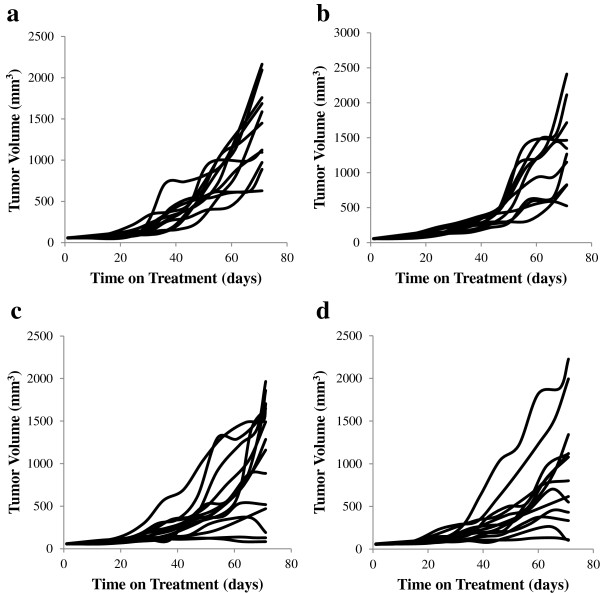
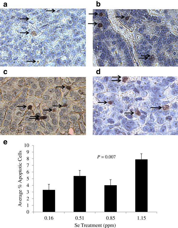
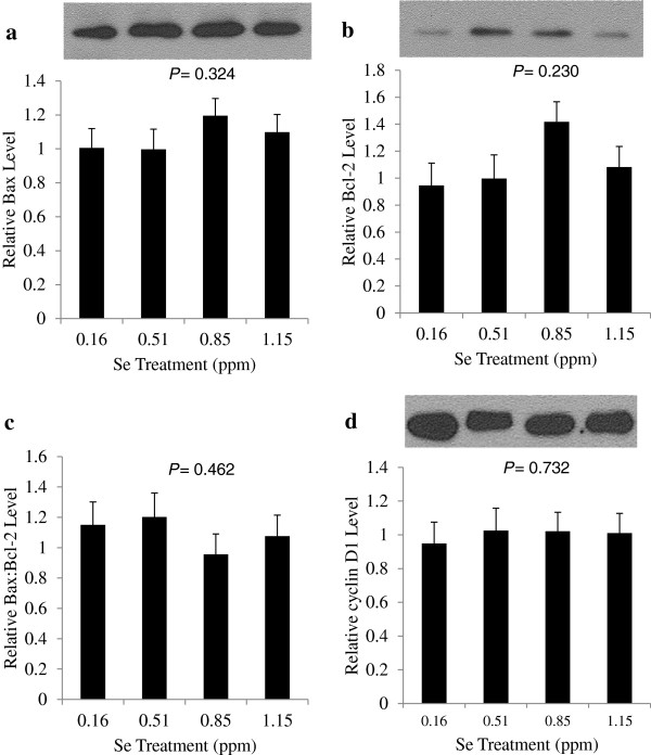
Methods: Four isonitrogenous diets with selenium levels of 0.16, 0.51, 0.85 and 1.15 ppm were formulated by mixing low- and high-selenium milk casein isolates with a rodent premix. MCF-7 cells were inoculated into the mammary fat pad of female BALB/c nude mice implanted with slow-release 17 β-estradiol pellets. Mice with palpable tumors were randomly assigned to one of the four diets for 10 weeks, during which time weekly tumor caliper measurements were conducted. Individual growth curves were fit with the Gompertz equation. Apoptotic cells and Bcl-2, Bax, and Cyclin D1 protein levels in tumors were determined.
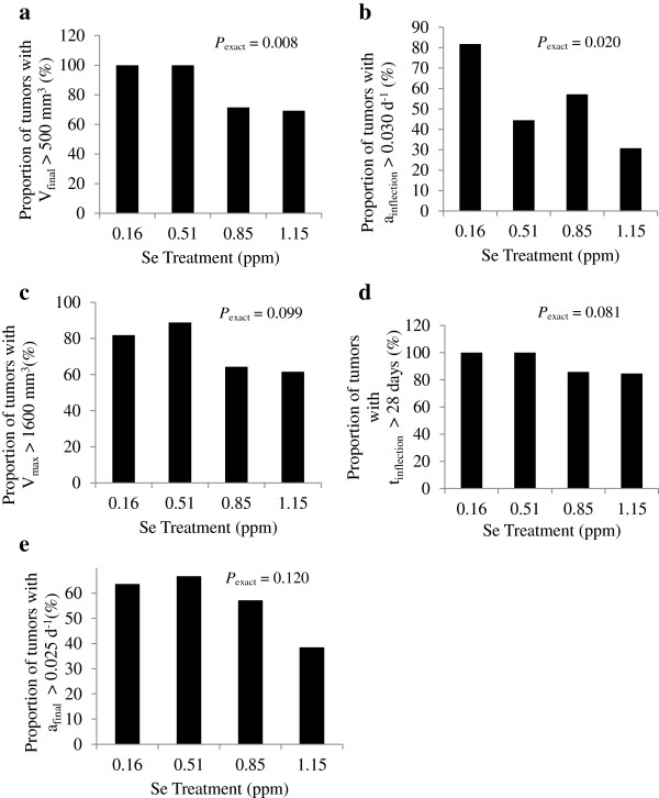
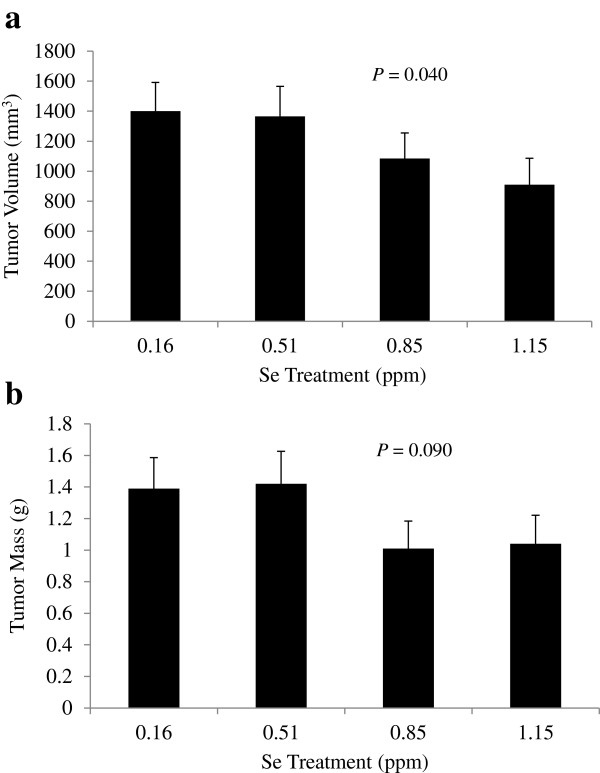
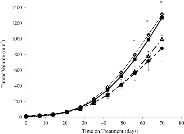
Results: There was a linear decrease in mean tumor volume at 70 days with increasing Se intake (P < 0.05), where final tumor volume decreased 35% between 0.16 and 1.15 ppm Se. There was a linear decrease in mean predicted tumor volume at 56, 63 and 70 days, and the number of tumors with a final volume above 500 mm3, with increasing Se intake (P < 0.05). This tumor volume effect was associated with a decrease in the proportion of tumors with a maximum growth rate above 0.03 day-1. The predicted maximum volume of tumors (Vmax) and the number of tumors with a large Vmax, were not affected by Se-casein. Final tumor mass, Bcl-2, Bax, and Cyclin D1 protein levels in tumors were not significantly affected by Se-casein. There was a significantly higher number of apoptotic cells in high-Se tumors as compared to low-Se tumors.
Conclusions: Taken together, these results suggest that turnover of cells in the tumor, but not its nutrient supply, were affected by dairy Se. We have shown that 1.1 ppm dietary Se from selenized casein can effectively reduce tumor progression in an MCF-7 xenograft breast cancer model. These results show promise for selenized milk protein as an effective supplement during chemotherapy.
Figures

Similar articles
-
Significance of amount and form of dietary selenium on blood, milk, and casein selenium concentrations in grazing cows.J Dairy Sci. 1999 Feb;82(2):429-37. doi: 10.3168/jds.S0022-0302(99)75249-5. J Dairy Sci. 1999. PMID: 10068964
-
The impact of organic vs. inorganic selenium on dairy goat productivity and expression of selected genes in milk somatic cells.J Dairy Res. 2019 Feb;86(1):48-54. doi: 10.1017/S0022029919000037. Epub 2019 Feb 13. J Dairy Res. 2019. PMID: 30758279
-
Dietary lutein inhibits mouse mammary tumor growth by regulating angiogenesis and apoptosis.Anticancer Res. 2003 Jul-Aug;23(4):3333-9. Anticancer Res. 2003. PMID: 12926072
-
Annonacin induces cell cycle-dependent growth arrest and apoptosis in estrogen receptor-α-related pathways in MCF-7 cells.J Ethnopharmacol. 2011 Oct 11;137(3):1283-90. doi: 10.1016/j.jep.2011.07.056. Epub 2011 Aug 4. J Ethnopharmacol. 2011. PMID: 21840388
-
Norcantharidin inhibits growth of human gallbladder carcinoma xenografted tumors in nude mice by inducing apoptosis and blocking the cell cycle in vivo.Hepatobiliary Pancreat Dis Int. 2010 Aug;9(4):414-22. Hepatobiliary Pancreat Dis Int. 2010. PMID: 20688607
Cited by
-
miR-148a and miR-17-5p synergistically regulate milk TAG synthesis via PPARGC1A and PPARA in goat mammary epithelial cells.RNA Biol. 2017 Mar 4;14(3):326-338. doi: 10.1080/15476286.2016.1276149. Epub 2017 Jan 17. RNA Biol. 2017. PMID: 28095188 Free PMC article.
-
Selenium-enriched plant foods: Selenium accumulation, speciation, and health functionality.Front Nutr. 2023 Feb 6;9:962312. doi: 10.3389/fnut.2022.962312. eCollection 2022. Front Nutr. 2023. PMID: 36815133 Free PMC article. Review.
-
Selenium-Based Novel Epigenetic Regulators Offer Effective Chemotherapeutic Alternative with Wider Safety Margins in Experimental Colorectal Cancer.Biol Trace Elem Res. 2022 Feb;200(2):635-646. doi: 10.1007/s12011-021-02659-5. Epub 2021 Mar 6. Biol Trace Elem Res. 2022. PMID: 33677818
-
Dual Functional Magnetic Nanoparticles Conjugated with Carbon Quantum Dots for Hyperthermia and Photodynamic Therapy for Cancer.Nanotheranostics. 2024 Apr 23;8(4):442-457. doi: 10.7150/ntno.91871. eCollection 2024. Nanotheranostics. 2024. PMID: 38961886 Free PMC article.
-
Micronutrients and Breast Cancer Progression: A Systematic Review.Nutrients. 2020 Nov 25;12(12):3613. doi: 10.3390/nu12123613. Nutrients. 2020. PMID: 33255538 Free PMC article.
References
-
- Clark LC, Combs GF, Turnbull BW, Slate EH, Chalker DK, Chow J, Davis LS, Glover RA, Graham GF, Gross EG, Krongrad A, Lesher JL, Park HK, Sanders BB, Smith CL, Taylor JR. Effect of selenium supplementation for cancer prevention in patients with carcinoma of the skin. J Am Med Assoc. 1996;13:1957–1963. doi: 10.1001/jama.1996.03540240035027. - DOI - PubMed
-
- Jiang C, Wang Z, Ganther H, Lu J. Distinct effects of methylseleninic acid versus selenite on apoptosis, cell cycle, and protein kinase pathways inDU145 human prostate cancer cells. Mol Cancer Ther. 2002;13:1059–1066. - PubMed
Publication types
MeSH terms
Substances
LinkOut - more resources
Full Text Sources
Other Literature Sources
Medical
Research Materials
