. 2013 Nov 7;503(7474):72-7.
doi: 10.1038/nature12630. Epub 2013 Oct 23.

SHANK3 overexpression causes manic-like behaviour with unique pharmacogenetic properties

Affiliations

SHANK3 overexpression causes manic-like behaviour with unique pharmacogenetic properties

Kihoon Han et al. Nature. .

Abstract

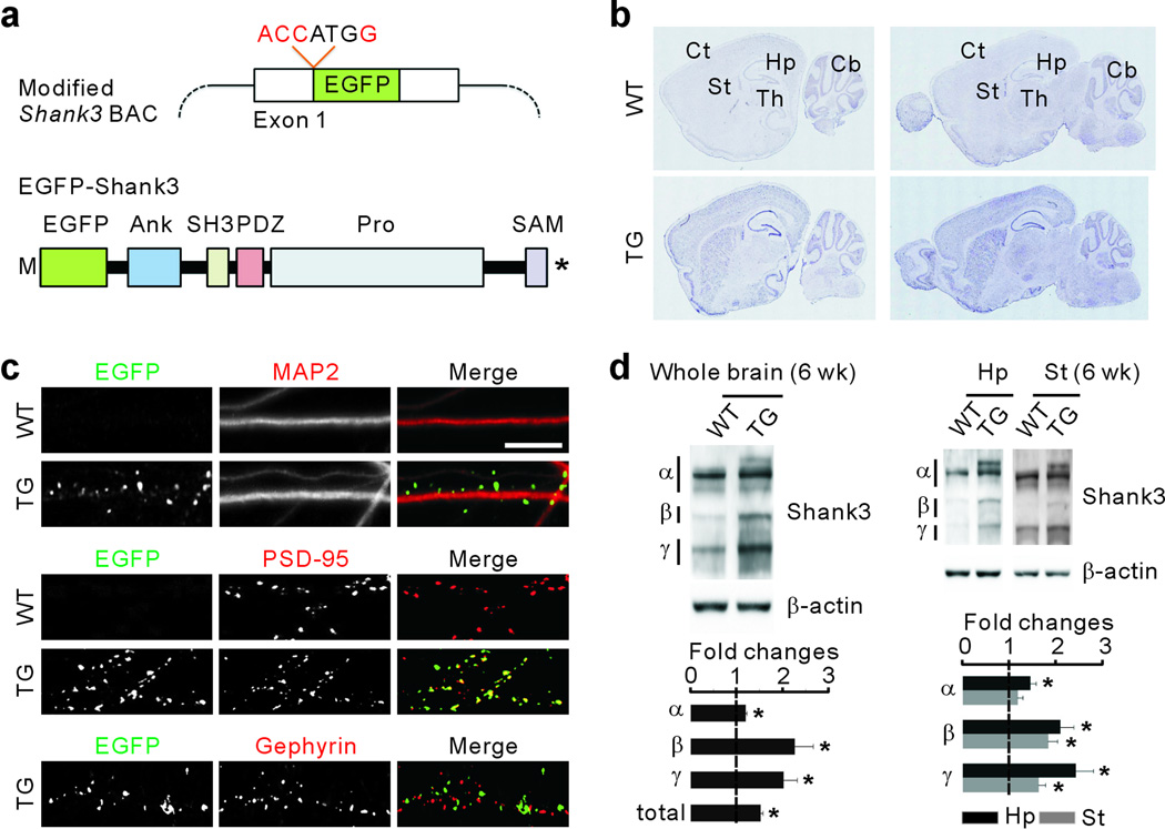
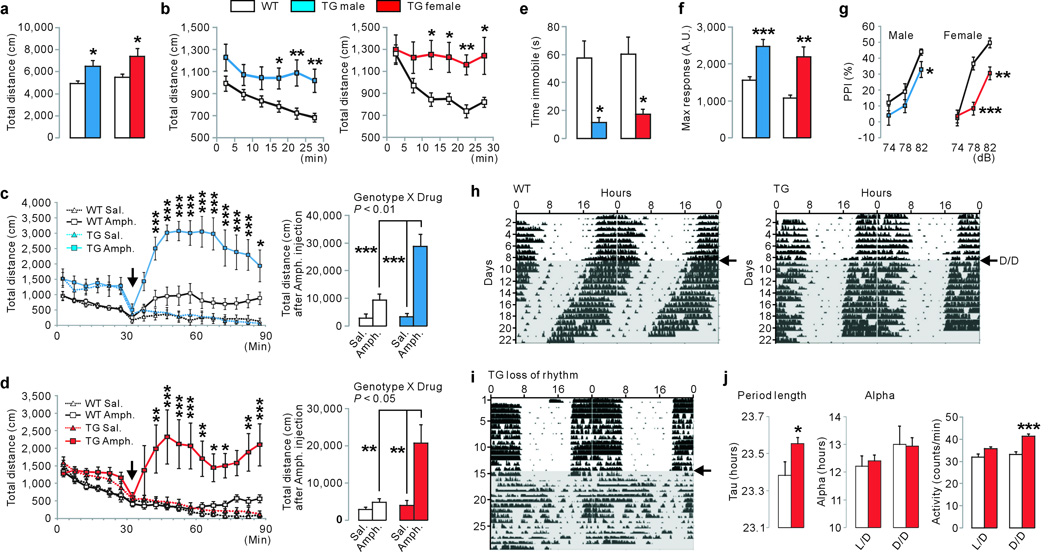
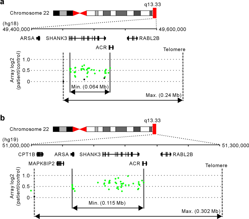
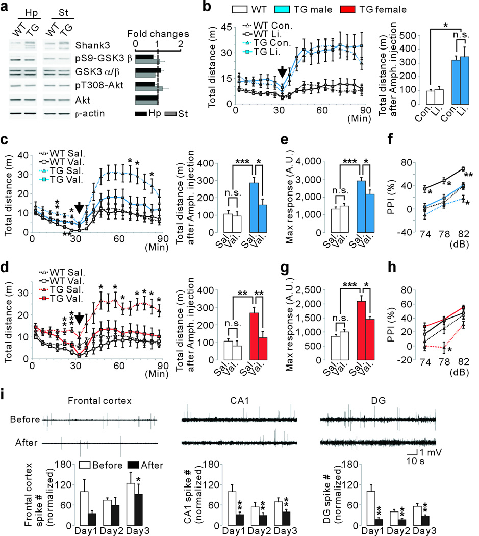
Mutations in SHANK3 and large duplications of the region spanning SHANK3 both cause a spectrum of neuropsychiatric disorders, indicating that proper SHANK3 dosage is critical for normal brain function. However, SHANK3 overexpression per se has not been established as a cause of human disorders because 22q13 duplications involve several genes. Here we report that Shank3 transgenic mice modelling a human SHANK3 duplication exhibit manic-like behaviour and seizures consistent with synaptic excitatory/inhibitory imbalance. We also identified two patients with hyperkinetic disorders carrying the smallest SHANK3-spanning duplications reported so far. These findings indicate that SHANK3 overexpression causes a hyperkinetic neuropsychiatric disorder. To probe the mechanism underlying the phenotype, we generated a Shank3 in vivo interactome and found that Shank3 directly interacts with the Arp2/3 complex to increase F-actin levels in Shank3 transgenic mice. The mood-stabilizing drug valproate, but not lithium, rescues the manic-like behaviour of Shank3 transgenic mice raising the possibility that this hyperkinetic disorder has a unique pharmacogenetic profile.


Figures

Figure 1
Figure 1. Characterization of EGFP-Shank3 expression in Shank3 transgenic mice
a, Diagrams show the modified Shank3 BAC and EGFP-Shank3. Kozak (ACCATGG) and EGFP-coding sequence were integrated into the start codon of Shank3. b, RNA in situ hybridization with a probe against EGFP detected EGFP-Shank3 in the brain. c, EGFP-Shank3 localizes to excitatory postsynaptic sites in cultured hippocampal neurons. MAP2 is a dendritic marker. Scale bar, 10 µm. d, Quantification of the fold changes of Shank3 (α, β and γ isoforms) in synaptosomal fraction of whole brain (n=4, biological replicates), hippocampus (Hp) or striatum (St) (n=6) from 6-week old (wk) mice. All data are presented as mean ± s.e.m. *P<0.05; unpaired two-tailed Student’s t-test.
Figure 2
Figure 2. Shank3 transgenic mice display manic-like behaviors
a, TG mice show increased locomotor activity in the open field test. b, TG mice did not habituate during the open field assay. c,d, TG mice are hypersensitive to amphetamine. After 30 min of basal activity, amphetamine (2 mg/kg) or saline was administered (arrow) and locomotor activity was monitored for 60 min. e, TG mice spend less time immobile in tail-suspension test. f, Increased acoustic (120 dB) startle response of TG mice. g, Abnormal PPI of TG mice. h–j, Abnormal circadian rhythms of TG mice. h, Wheel running actograms of WT and TG female mice. After 8 days of light/dark (L/D) cycle, animals were released into constant darkness (arrow, D/D) for 2 weeks. i, Among 20 TG mice tested, 3 displayed complete loss of rhythm during D/D. j, TG mice showed increased period length and activity count, but normal alpha, compared to WT mice. All data are presented as mean ± s.e.m. *P<0.05; **P<0.01; ***P<0.001. Statistical analyses for behavioral assays are in Supplementary Table 1.
Figure 3
Figure 3. Individuals with SHANK3 duplications have hyperkinetic disorders
a, Array plot of an exon-targeted chromosome microarray analysis on DNA from an 11-year old female with ADHD, seizures and aberrant behaviors. Black dots indicate probes with normal copy number, while green dots indicate copy number gain. Solid and dotted lines define the minimum and maximum expected boundaries of the duplication, respectively. b, Array plot of the 35-year old male with bipolar disorder and epilepsy.
Figure 4
Figure 4. Abnormal EEG and altered synaptic excitatory/inhibitory balance of Shank3 transgenic mice
a, Representative EEG traces from WT (n=5) and TG (n=10) mice. TG mice showed prolonged hyperexcitability discharges and electrographic seizure in all recorded regions. The frequency of epileptiform spikes (dentate gyrus) and electrographic seizure were significantly increased in TG mice. b, Increased VGLUT1-positive PSD-95 (n=28, three independent experiments) and decreased VGAT-positive Gephyrin (n=25) puncta density in cultured hippocampal pyramidal neurons from TG mice. Scale bar, 10 µm. c, Frequency, but not amplitude and decay, of mIPSC was decreased in CA1 pyramidal neurons of TG mice (WT n=14, TG n=18). d, Amplitude, but not frequency and decay, of sEPSC was increased in TG neurons (n=19). e, AMPA/NMDA ratio was normal in TG neurons (n=9). All data are presented as mean ± s.e.m. *P<0.05; **P<0.01. Statistical analyses for these data are in Supplementary Table 2 and 3.
Figure 5
Figure 5. Shank3 interacts directly with Arp2/3 complex to increase F-actin levels in Shank3 transgenic mice
a, Increased synaptic F-actin in cultured hippocampal pyramidal neurons from TG mice (n=29, three independent experiments). Scale bar, 10 µm. b, Actin cytoskeleton-related sub-network of Shank3 interactome. c, Increased ARPC2 cluster size in TG pyramidal neurons (n=20). d, Diagram shows the proposed role of Shank3 as a platform for F-actin regulating proteins. e, Increased co-localization of ARPC2 and WASF1 in TG pyramidal neurons (n=22). f, Golgi-staining of CA1 pyramidal neurons shows more dendritic spines in TG mice (n=40 neurons from 3 animals per genotype). g,h, Excitatory and inhibitory synaptic distributions of Mena (g) and Profilin2 (h) in cultured CA1 pyramidal neurons. Yellow arrowheads indicate protein puncta co-localized with corresponding synaptic markers. i, Quantification of (g) and (h). Density of VGAT-positive Mena puncta is decreased in TG neurons (n=18). VGLUT1-positive Profilin2 puncta intensity is increased in TG neurons (n=13), while VGAT-positive Mena and Profilin2 puncta intensity are decreased in TG neurons. All data are presented as mean ± s.e.m. *P<0.05; **P<0.01.
Figure 6
Figure 6. Valproate, but not lithium, rescues manic-like behaviors of Shank3 transgenic mice
a, Basal activities of GSK-3β and Akt in the hippocampus and striatum of TG mice (10-week old, n=7) are normal. b, Amphetamine-sensitivity of male TG mice was not rescued by lithium. c,d, Basal locomotor activity and amphetamine-sensitivity of TG mice were rescued by valproate. Basal activity of WT male mice was decreased by valproate during one 5-min time bin. e–h Acoustic startle response (e,g) and PPI (f,h) of TG mice were rescued by valproate. Valproate increased PPI of WT male mice. i, Rescue of abnormal EEG in TG mice by valproate. During the three consecutive days of tests, EEG was recorded from the three brain regions of TG mice (n=7) for one hour before and after valproate injection. Representative EEG traces measured on day 1 are shown. The number of epileptiform spikes was quantified and normalized to the baseline values before treatment of day 1. All data are presented as mean ± s.e.m. *P<0.05; **P<0.01; ***P<0.001.

Comment in

References

    1. Sudhof TC. Neuroligins and neurexins link synaptic function to cognitive disease. Nature. 2008;455:903–911. - PMC - PubMed
    1. Zoghbi HY. Postnatal neurodevelopmental disorders: meeting at the synapse? Science. 2003;302:826–830. - PubMed
    1. Bourgeron T. A synaptic trek to autism. Curr Opin Neurobiol. 2009;19:231–234. - PubMed
    1. Ting JT, Peca J, Feng G. Functional consequences of mutations in postsynaptic scaffolding proteins and relevance to psychiatric disorders. Annu Rev Neurosci. 2012;35:49–71. - PubMed
    1. Sheng M, Kim E. The Shank family of scaffold proteins. J Cell Sci. 2000;113(Pt 11):1851–1856. - PubMed

Publication types

MeSH terms