. 2013:4:2660.
doi: 10.1038/ncomms3660.

Visualization and targeted disruption of protein interactions in living cells

Affiliations

Visualization and targeted disruption of protein interactions in living cells

Henry D Herce et al. Nat Commun. 2013.

Abstract

Protein-protein interactions are the basis of all processes in living cells, but most studies of these interactions rely on biochemical in vitro assays. Here we present a simple and versatile fluorescent-three-hybrid (F3H) strategy to visualize and target protein-protein interactions. A high-affinity nanobody anchors a GFP-fusion protein of interest at a defined cellular structure and the enrichment of red-labelled interacting proteins is measured at these sites. With this approach, we visualize the p53-HDM2 interaction in living cells and directly monitor the disruption of this interaction by Nutlin 3, a drug developed to boost p53 activity in cancer therapy. We further use this approach to develop a cell-permeable vector that releases a highly specific peptide disrupting the p53 and HDM2 interaction. The availability of multiple anchor sites and the simple optical readout of this nanobody-based capture assay enable systematic and versatile analyses of protein-protein interactions in practically any cell type and species.


Figures

Figure 1
Figure 1. Strategy for visualizing protein interactions in living cells and outline of a fundamental biological application.
(a) Schematic representation of the fluorescence three-hybrid (F3H) to study protein–protein interactions. A GFP binder protein (GBP) is attached to a protein (LP) that accumulates at a well-defined location within the cell. This complex recruits to that particular location GFP-tagged proteins (GFP-P1). If the protein P1 interacts with a second protein P2, labelled with a different fluorescent marker, the interaction can be immediately visualized using fluorescent microscopy. (b) Schematic representation of the interaction between p53 and HDM2/X and its central role in cellular regulation and genome preservation.
Figure 2
Figure 2. Targeted recruitment of GFP-tagged proteins allows visualization and quantification of protein–protein interactions in live cells.
Re-localization of GFP-tagged proteins (tethered; immobilized; recruited) to the Lac operator using the GBP–LacI. Confocal microscopy images of non-interacting GFP and mCherry are shown and interacting GFP–p53 (pNeG-p53(NTD)) and mCh–HDM2 (pCAG-Ch-HDM2(NTD)) proteins in live cells co-transfected with and without the GBP–LacI. In the first row, a schematic representation of the transfected constructs and the respective protein distribution is shown, in the second row, a confocal image of the GFP-tagged protein, in the third row, an image of the mCherry-tagged protein, in the fourth row, the overlay of the two previous channels and in the last row, the derivative of the Pearson’s correlation coefficient between the GFP image and the mCherry image along the dotted line drawn in the confocal images. In columns (ad) representative images of cells transiently transfected with the constructs indicated in the respective schemes above are shown. Scale bar, 5 μm.
Figure 3
Figure 3. Visualization of targeted disruption of protein interactions in live cells.
BHK cells containing a stably integrated LacO array were transiently transfected with pNeG-p53(NTD), pCAG-Ch-HDM2(NTD) and GBP–LacI. In (a) is shown a schematic representation of the re-localization of the GFP–p53 to the LacO array, its interaction with mCh–HDM2 and the disruption of this interaction mediated by Nutlin 3. (b) Live cell confocal microscopy images showing the disruption kinetics of the interaction between HDM2 and p53 mediated by Nutlin 3 at 0 (DMSO, control), 2, 5 and 10 μM. (c) Time lapse quantification of the relative binding of p53 to HDM2. Higher concentrations of Nutlin 3 resulted in faster disruption of the interaction between the proteins. In Supplementary Fig. S1 are shown the kinetic traces, the mean and the s.e. of the interaction disruption mediated by Nutlin 3 at 5 μM obtained from five repetitions showing the reproducibility of the individual traces. Scale bar, 5 μm.
Figure 4
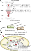
Figure 4. Rationally designed in vivo cleavable cell-permeable peptide to inhibit and disrupt the binding between p53 and HDM2.
(a) Short sequence from p53 responsible for the binding between p53 and HDM2. Below this sequence the main domains of p53 and HDM2 are depicted along with a short peptide (N8A) that has been shown in vitro to have higher affinity for HDM2 than p53. (b) The N8A peptide is not able to reach the interior of living cells. Therefore, it was coupled by a disulphide bond to a cell-penetrating peptide (TAT) that is capable of transporting it into living cells. (c) The design is based on a delivery model in which (I) the TAT peptide transports the N8A peptide into the cell, (II) in the cytosol the disulphide bridge is cleaved releasing the N8A peptide from the TAT peptide, (III) the peptide inhibitor is free to diffuse throughout the nucleus and the cytosol inhibiting the binding between p53 and HDM2, (IV) while the TAT peptide accumulates preferentially at the cytosol and the nucleolus.
Figure 5
Figure 5. A rationally designed in vivo cleavable cell-permeable peptide that inhibits and disrupts the binding between p53 and HDM2.
The peptide was tested in several mammalian cell lines (ad). Starting from hamster cell lines containing stably integrated Lac operator (LacO) DNA arrays, the system is expanded onto application on any mouse cell exploiting the natural occurrence of large regions around centromeres naturally containing arrays of major satellite DNA sequences rich in methylated cytosines and, hence, accumulating methyl cytosine-binding domain (MBD) proteins. On a next step, the system is expanded to any cell containing a nuclear lamina composed of lamin intermediate filament proteins. Finally, the system is transferred to the cytosol making use of targeting it to the centriole via the centrin protein. In the first column, a cartoon representation of the protein interactions and inhibition at each recruitment site within the cell is shown. In the second column, a confocal image of a representative live cell of the GFP channel is shown followed by the mCherry channel in the third column, a transmission light image in the fourth column and in the last column the overlay of all the channels. In each row are shown the images before and after adding the N8A peptide to a final concentration of 8 μM. An amplified image of the recruitment site is shown below each microscopic image. (a) Recruitment of GFP-HDM2 to the LacO array in hamster BHK cells. Cells transiently transfected with pNeG-HDM2(NTD), pCAG-Ch-p53(NTD) and GBP–LacI. (b) Recruitment of GFP-HDM2 to major satellite pericentric repeats rich in MBD proteins in mouse C2C12 cells. Cells transiently transfected with pNeG-HDM2(NTD), pCAG-Ch-p53(NTD) and GBP-MBD (c) Recruitment of GFP-HDM2 to the nuclear lamina in human HeLa cells. Cells transiently transfected with pNeG-HDM2(NTD), pCAG-Ch-p53(NTD) and GBP-Lamin B1. (d) Recruitment of p53-GFP to the centriole in HeLa cells. Cells transiently transfected with p53-GFP, pCAG-Ch-HDM2(NTD) and GBP-Centrin2. Scale bar, 5 μm.

References

    1. Fields S. & Song O. A novel genetic system to detect protein-protein interactions. Nature 340, 245–246 (1989). - PubMed
    1. Cremazy F. G. et al. Imaging in situ protein-DNA interactions in the cell nucleus using FRET-FLIM. Exp. Cell. Res. 309, 390–396 (2005). - PubMed
    1. Tsuganezawa K. et al. A fluorescent-based high-throughput screening assay for small molecules that inhibit the interaction of MdmX with p53. J. Biomol. Screen. 18, 191–198 (2013). - PubMed
    1. Kirchhofer A. et al. Modulation of protein properties in living cells using nanobodies. Nat. Struct. Mol. Biol. 17, 133–138 (2010). - PubMed
    1. Montes de Oca Luna R., Wagner D. S. & Lozano G. Rescue of early embryonic lethality in mdm2-deficient mice by deletion of p53. Nature 378, 203–206 (1995). - PubMed

Publication types

MeSH terms