. 2013 Oct 23;5(208):208ra149.
doi: 10.1126/scitranslmed.3007529.

Targeting RNA foci in iPSC-derived motor neurons from ALS patients with a C9ORF72 repeat expansion

Affiliations

Targeting RNA foci in iPSC-derived motor neurons from ALS patients with a C9ORF72 repeat expansion

Dhruv Sareen et al. Sci Transl Med. .

Abstract

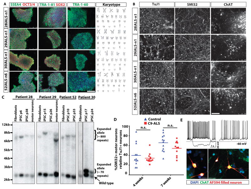
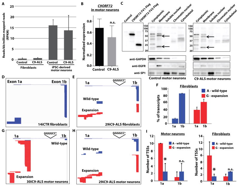
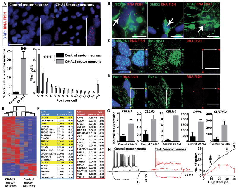
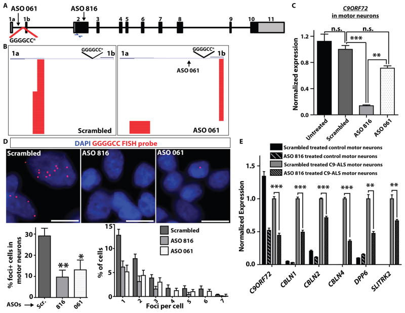
Amyotrophic lateral sclerosis (ALS) is a severe neurodegenerative condition characterized by loss of motor neurons in the brain and spinal cord. Expansions of a hexanucleotide repeat (GGGGCC) in the noncoding region of the C9ORF72 gene are the most common cause of the familial form of ALS (C9-ALS), as well as frontotemporal lobar degeneration and other neurological diseases. How the repeat expansion causes disease remains unclear, with both loss of function (haploinsufficiency) and gain of function (either toxic RNA or protein products) proposed. We report a cellular model of C9-ALS with motor neurons differentiated from induced pluripotent stem cells (iPSCs) derived from ALS patients carrying the C9ORF72 repeat expansion. No significant loss of C9ORF72 expression was observed, and knockdown of the transcript was not toxic to cultured human motor neurons. Transcription of the repeat was increased, leading to accumulation of GGGGCC repeat-containing RNA foci selectively in C9-ALS iPSC-derived motor neurons. Repeat-containing RNA foci colocalized with hnRNPA1 and Pur-α, suggesting that they may be able to alter RNA metabolism. C9-ALS motor neurons showed altered expression of genes involved in membrane excitability including DPP6, and demonstrated a diminished capacity to fire continuous spikes upon depolarization compared to control motor neurons. Antisense oligonucleotides targeting the C9ORF72 transcript suppressed RNA foci formation and reversed gene expression alterations in C9-ALS motor neurons. These data show that patient-derived motor neurons can be used to delineate pathogenic events in ALS.


Conflict of interest statement

Competing interests: FB and FR are employees of Isis Pharmaceuticals and hold stock options in the company. FB serves on the scientific advisory board of the Experimental Therapeutic Centre, Singapore. FB has submitted patents related to this work regarding the design and use of antisense oligonucleotides targeting the C9ORF72 transcript.

Figures

Fig. 1
Fig. 1. Generation of C9ORF72 ALS patient-derived iPSCs and motor neurons
(A) Pluripotency marker expression in iPSC lines derived from four ALS patients with a C9ORF72 repeat expansion. Immunostaining shows expression of embryonic and pluripotency stem cell surface antigens, SSEA4, TRA-1-60, TRA-1-81; and nuclear pluripotency markers OCT3/4 and SOX2. Karyotypes of the four C9ORF72 patient iPSC lines are shown on the right. (B) Efficient production of motor neurons from ALS C9ORF72 patient-derived iPSCs demonstrated by SMI32+ and ChAT+ immunostaining. TuJ1 is a pan-neuronal marker used to assess for total production of neurons. (C) Examination of hexanucleotide repeat lengths in fibroblasts, iPSCs and motor neuron cultures by Southern blot analysis, showing somatic instability of the repeat with both expansion (iPSCs in lines 28i,29i,52i) and contraction (motor neurons in iPSC line derived from patient 29). Passage numbers for iPSCs are shown (p). (D) Motor neuron survival over time was not altered in the iPSC-derived motor neuron cultures derived from four individual C9ORF72 ALS (C9-ALS) patients versus four control subjects (n=3 independent experiments) (1-way ANOVA, Tukey’s Multiple Comparison Test; Control vs. ALS, 95% confidence interval of difference, 4 weeks: −12.8 to 24.15 and 7 weeks: −7.36 to 30.42). Motor neuron counts are represented as a ratio of motor neuron (SMI32+) to pan-neuronal (TuJ1+) populations. (E) Properties of functional motor neurons were observed when iPSCs were differentiated into motor neurons (representative motor neuron from control iPSC line 83i control shown at day 69 of differentiation). Current injections of −10 pA and +5 pA (top left). Resting potential of the motor neuron was −60mV with 2.6 GigaOhm input resistance. Spontaneous activity of the same motor neuron (top right). Representative images confirming motor neuron identity of recorded neurons (bottom panels). Neurons filled with Alexa594 dye (red) during live electrophysiological recordings are shown post-fixation and counter-stained with motor neuron marker ChAT (green) confirming their identity.
Fig. 2
Fig. 2. The hexanucleotide expansion does not alter expression of C9ORF72 but alters upstream exon utilization to promote transcription of the repeats
(A) Reads per kilobase per million mapped reads (RPKM) from RNA sequencing of motor neurons differentiated from iPSCs derived from control individuals (n=4 independent subjects) and ALS patients with the C9ORF72 repeat expansions (C9-ALS, n=4 independent subjects) and fibroblasts, for all annotated transcripts from the C9ORF72 gene. Overall expression of C9ORF72 transcripts was not different between controls and C9ORF72 expansion carriers, with motor neuron cultures showing higher RPKM values than fibroblasts. (B) Quantitative RT-PCR in iPSC-derived motor neuron cultures from four control subjects and four C9ORF72 expansion carriers using primers in exon 2 (common to all C9ORF72 transcript variants), confirming equivalent expression of the C9ORF72 gene. Data are represented as mean ± SEM from n=3 independent experiments. n.s., not significant using student’s t-test. (C) Western blot of C9ORF72 protein in cellular fractions from iPSC-derived motor neuron cultures showing two bands in the membrane fraction running slightly smaller than the sizes of overexpressed long and short isoforms due to the presence of a Flag tag on these constructs. Representative blots showing similar levels of protein were observed in control (83iCTR) and C9ORF72 expansion (28iC9-ALS) motor neuron cultures indicating that the presence of the repeat did not alter overall protein levels. Data are representative of n=3 independent experiments. (D) Representative sequence alignments of upstream exon sequences (exons 1a or 1b) from 5′ RACE analysis of C9ORF72 transcripts in fibroblasts from a control subject (14iCTR). Stacked horizontal bars represent individual transcripts, with 100 transcripts sequenced for each sample. In control cells, transcripts containing exon 1b were most frequent, with little variability in transcriptional start sites for either exon 1a or exon 1b. (E) Alignment of 5′ RACE transcripts from fibroblasts from a C9ORF72 expansion patient (29iC9-ALS). Sequences derived from the wild-type or expansion allele were determined using SNP rs10757668 upstream of the RACE primer in exon 2. Expression of the wild-type allele (blue) was similar to control fibroblasts, whereas the expansion allele (red) showed more frequent usage of exon 1a and more variability in the transcriptional start site. (F) Percentage of transcripts containing exon 1a or 1b in C9ORF72 patient fibroblasts (29i and 30i) from the wild-type (A allele, blue) or expansion (G allele, red), showing an increase in the percentage of transcripts containing exon 1a derived from the expansion allele. *p<0.05, t-test, % of transcripts, wild-type vs. mutant allele. (G, H) Alignment of 5′ RACE from iPSC-derived motor neurons from two different C9ORF72 expansion ALS patients (30iC9-ALS and 29iC9-ALS), showing enhanced expression of the repeat (containing exon 1a), and variability in transcriptional start site usage by the mutant allele. (I) Comparison of the number of transcriptional start sites (TSSs) observed in fibroblasts from C9ORF72 patients 29i and 30i (right panel) and iPSC-derived motor neurons (left panel). The expansion allele (G, red) showed more TSSs for exon 1a transcripts than the wild-type allele (A, blue), whereas the number of TSSs for exon 1b was similar between the two alleles. *p<0.05, t-test, # of TSSs, wild-type (A) versus mutant (G).
Fig. 3
Fig. 3. iPSC-derived motor neuron cultures from C9-ALS patients develop GGGGCC RNA foci that bind to Pur-α and hnRNPA1
(A) Representative images of fluorescence in situ hybridization (FISH) with an antisense probe to the GGGGCC repeat in iPSC-derived motor neuron cultures (lines 83iCTR and 52iC9-ALS). RNA foci were present in cells from all C9-ALS patients, but not in motor neuron cultures from control subjects. RNA foci were predominantly nuclear, but occasionally found in the cytoplasm as well. Scale bar = 25 μm. Histogram below shows the number of foci per cell. **p<0.01 unpaired t-test (two-tailed). ***control (all four subjects) vs. C9-ALS (all four subjects) p<0.0001 Mann Whitney test. (B) Representative images showing co-staining of GGGGCC FISH and markers of different cell types in iPSC-derived motor neuron cultures. RNA foci were present in the nuclei of neuronal precursors (nestin positive; line 29iC9-ALS shown), motor neurons (SMI32 positive; line 28iC9-ALS shown), and astroglial cells (GFAP positive; line 52iC9-ALS shown). Scale bar = 10 μm. (C, D) Representative images showing co-staining of GGGGCC FISH with RNA binding proteins. Co-localization of GGGGCC foci with hnRNPA1 and Pur-α was observed by confocal imaging. White arrows point to foci that stained for both RNA foci and hnRNPA1 or Pur-α in the same focal plane. Adjacent panels show the y-z and x-z axes confirming colocalization in 3 dimensions. Scale bar = 10 μm. C, line 52iC9-ALS; D, line 28iC9-ALS. (E) Heat map showing hierarchical clustering of differentially expressed genes identified by RNA-seq of iPSC-derived motor neuron cultures between four different C9-ALS patients (n=4; lines 28i, 29i, 30i, 52i) and four control subjects (n=4; lines 00i, 03i, 14i, 83i) with p< 0.05. The arrow indicates motor neuron cultures from the subject with the fewest GGGGCC repeats (~70 in line 30iC9-ALS), which clustered farthest from the three lines with larger repeats (~800). (F) Gene list of 20 highest upregulated (blue) and downregulated (red) genes in C9-ALS patient iPSC-derived motor neuron cultures versus controls. Genes highlighted in yellow include DPP6, implicated in prior ALS GWAS studies, and three members of the cerebellin family (CBLN1, , and 4). (G) qRT-PCR validation of differentially expressed genes highlighted in (F), all p<0.05, CTR (n=4 subjects) vs C9-ALS (n=4 subjects), t-test. (H) C9-ALS iPSC-derived motor neuron cultures are less excitable (n=184) than control iPSC-derived motor neuron cultures (n=137). Recordings were performed on motor neuron cultures that were between days 66–79 of differentiation. Representative traces are shown in response to current injections of −10, 0 and 10 pA into motor neurons derived from a control subject (black, iPSC line 83i) or C9-ALS patient (red, iPSC line 28i). Mean number of action potentials elicited as a function of current injection for control (black) and C9-ALS (red) cultured motor neurons. The graph (right) shows the number of spikes fired at different levels of current injection, with reduced numbers of spikes fired in C9-ALS iPSC-derived motor neurons compared to control iPSC-derived motor neurons (CTR, iPSC lines 14i and 83i, C9-ALS iPSC lines 28i and 52i; n=2 independent experiments). Resting potential and input resistance of the motor neurons is −68.5 mV with 3.4 GigaOhm for the control and −61.5 mV with 3.2 GigaOhm for the C9-ALS motor neurons. *p<0.05; **p<0.01; ***p<0.001; C9-ALS vs. control, unpaired t-test, two sample, unequal variance.
Fig. 4
Fig. 4. Knockdown of C9ORF72 with antisense oligonucleotides suppresses RNA foci in ALS motor neurons
(A) Schematic diagram of C9ORF72 gene structure, showing the location of antisense oligonucleotides (ASO) 816 and 061, and primers for assaying C9ORF72 expression in exon 2. (B) Quantitative RT-PCR for total C9ORF72 in iPSC-derived motor neurons (C9-ALS lines 28iC9-ALS and 52iC9-ALS) treated with ASOs 816 and 061. ASO816, which targets the first coding exon common to all C9ORF72 isoforms, knocked down overall C9ORF72 levels by ~90%. ASO061 had a partial effect on total transcript levels. (1-way ANOVA Tukey’s Multiple Comparison Test, 95% CI of diff; untreated vs. scrambled (n.s.): −0.1713 to 0.4167; scrambled vs. ASO061 (n.s.): −0.006394 to 0.5816; scrambled vs. ASO816 (***): − 0.5671 to 1.155; ASO816 vs. ASO061 (**.):−0.8675 to −0.2796). n.s. = not significant. Data are represented as mean ± SEM from n=3 independent experiments. (C) 5′ RACE analysis (line 52iC9-ALS shown) to analyze 5′ exon utilization after treatment with either scrambled ASO (left) or ASO061 (right). While ASO061 did not alter total C9ORF72 transcript levels, it suppressed exon 1a containing transcripts, with a relative increase in exon 1b containing transcripts. (D) Representative images of RNA FISH on C9-ALS patient motor neuron cultures (lines 29iC9-ALS and 52iC9-ALS) treated with different ASOs (scrambled – “Scr.”, 816, 061) showing a marked suppression of RNA foci in ASO-treated cells. Scale bar = 10 μm. Graphs below show quantitation of the percentage of cells with foci (left), and a histogram showing the breakdown by foci per cell (right). ** = p<0.01; * = p<0.05; unpaired t-test (two-tailed). (E) Quantitative RT-PCR for genes that showed aberrant upregulation in C9-ALS iPSC-derived motor neuron cultures (DPP6, CBLN1, CBLN2, CBLN4 and SLITRK2) after treatment with scrambled ASO or ASO816 (lines 14iCTR and 52iC9-ALS shown). Error bars are mean ± sem. ***p<0.001; **p<0.01; unpaired t-test (two-tailed), scrambled vs. ASO816.

Similar articles

Cited by

References

    1. Lomen-Hoerth C, Anderson T, Miller B. The overlap of amyotrophic lateral sclerosis and frontotemporal dementia. Neurology. 2002 Oct 8;59:1077. - PubMed
    1. Chen-Plotkin AS, Lee VM, Trojanowski JQ. TAR DNA-binding protein 43 in neurodegenerative disease. Nat Rev Neurol. 2010 Apr;6:211. - PMC - PubMed
    1. Ravits J, et al. Deciphering amyotrophic lateral sclerosis: what phenotype, neuropathology and genetics are telling us about pathogenesis. Amyotrophic lateral sclerosis & frontotemporal degeneration. 2013 May;14(Suppl 1):5. - PMC - PubMed
    1. Seeley WW. Selective functional, regional, and neuronal vulnerability in frontotemporal dementia. Curr Opin Neurol. 2008 Dec;21:701. - PMC - PubMed
    1. Neumann M, et al. Ubiquitinated TDP-43 in frontotemporal lobar degeneration and amyotrophic lateral sclerosis. Science. 2006 Oct 6;314:130. - PubMed

Publication types

MeSH terms