. 2013 Nov 26;110(48):E4571-80.
doi: 10.1073/pnas.1311669110. Epub 2013 Oct 23.

IFI16 senses DNA forms of the lentiviral replication cycle and controls HIV-1 replication

Affiliations

IFI16 senses DNA forms of the lentiviral replication cycle and controls HIV-1 replication

Martin R Jakobsen et al. Proc Natl Acad Sci U S A. .

Erratum in

  • Proc Natl Acad Sci U S A. 2013 Nov 26;110(48):19651. Jin, Tengchuan [corrected to Tengchuan, Jin]

Abstract

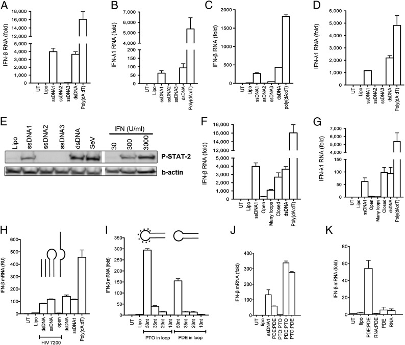
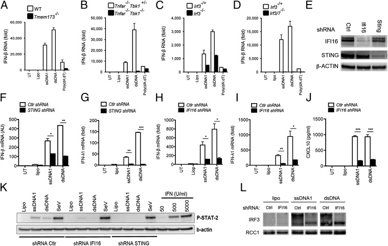
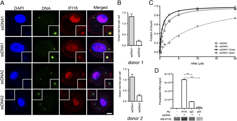
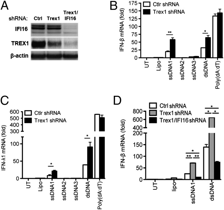
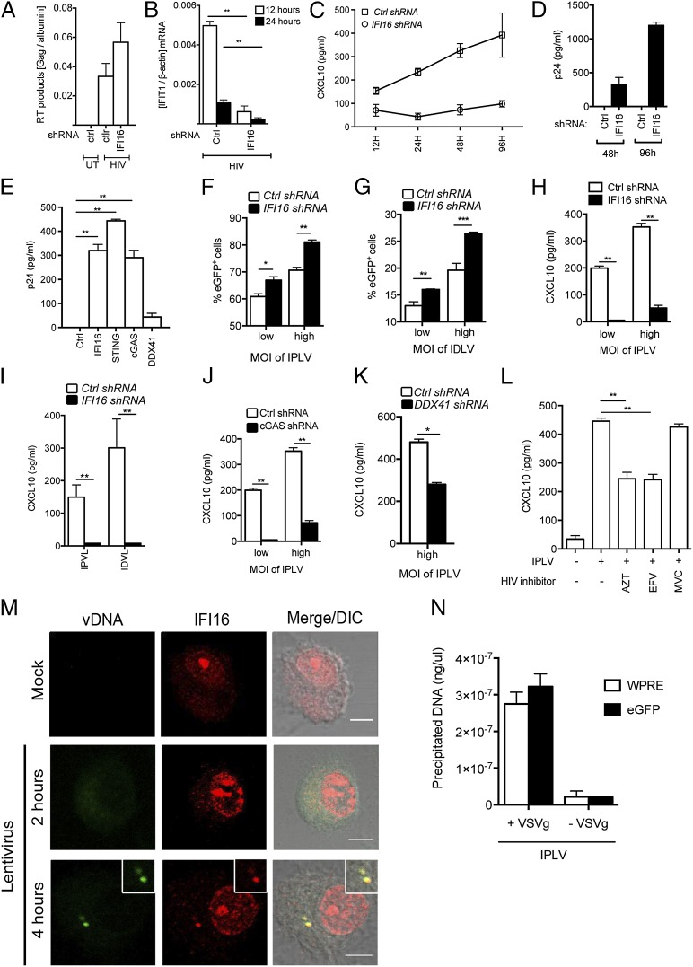
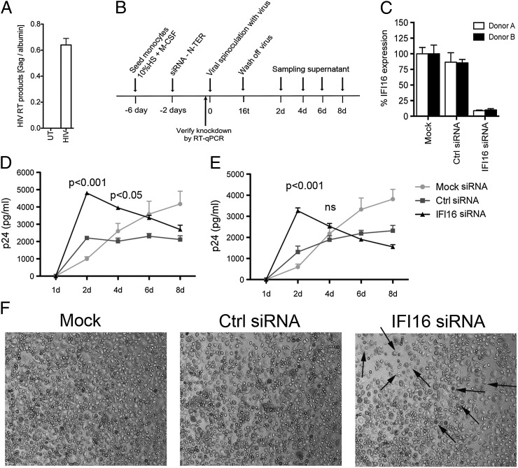
Replication of lentiviruses generates different DNA forms, including RNA:DNA hybrids, ssDNA, and dsDNA. Nucleic acids stimulate innate immune responses, and pattern recognition receptors detecting dsDNA have been identified. However, sensors for ssDNA have not been reported, and the ability of RNA:DNA hybrids to stimulate innate immune responses is controversial. Using ssDNAs derived from HIV-1 proviral DNA, we report that this DNA form potently induces the expression of IFNs in primary human macrophages. This response was stimulated by stem regions in the DNA structure and was dependent on IFN-inducible protein 16 (IFI16), which bound immunostimulatory DNA directly and activated the stimulator of IFN genes -TANK-binding kinase 1 - IFN regulatory factors 3/7 (STING-TBK1-IRF3/7) pathway. Importantly, IFI16 colocalized and associated with lentiviral DNA in the cytoplasm in macrophages, and IFI16 knockdown in this cell type augmented lentiviral transduction and also HIV-1 replication. Thus, IFI16 is a sensor for DNA forms produced during the lentiviral replication cycle and regulates HIV-1 replication in macrophages.

Keywords: DNA sensing; antiviral defense; innate immunity.


Conflict of interest statement

The authors declare no conflict of interest.

Figures

Fig. 1.
Fig. 1.
ssDNA induces type I and III IFN responses in human macrophages. (A–D) hMDMs (A and B; n = 4) and THP1 cells (C and D; n = 5) were transfected with three ssDNA oligo nucleotides (ssDNA1–3) derived from HIV-1, dsDNA, or poly(dA:dT), respectively. RNA was collected after 6 h of incubation and analyzed for levels of IFN-β (A and C) and IFN-λ1 (B and D) mRNA by RT-qPCR. (E) Supernatants from cells treated as in C and D for 20 h were used to stimulate parental THP1 cells for 50 min. Cell lysates were analyzed for STAT2 phosphorylation WB. Recombinant IFN-α was used as standard, and Sendia virus (SeV) was used as control. (F and G) Variants of ssDNA1 (open, many loops, and closed) were transfected into hMDMs, and RNA was isolated and analyzed for IFN-β (F) and IFN-λ1 (G) mRNA expression (n = 4). (HK) THP1 cells were transfected with the indicated DNA oligos. RNA was collected after 6 h of incubation and was analyzed for IFN-β mRNA expression by RT-qPCR (n = 2–4). Asterisks in I indicate stem-loop DNA with PTO in the loop region. All data are presented as a representative dataset from one of multiple independent experiments performed in duplicate (mean ± SEM). Lipo, lipofectamine; UT, untreated.
Fig. 2.
Fig. 2.
HIV-derived ssDNA induces IFN responses through a pathway dependent on IFI16, STING, TBK1, and IRF3/7. (AD) BMDMs from mice with homozygous deletions of the genes encoding (A) Tmem173 (i.e., STING) (n = 3), (B) TBK1 (n = 2), (C) IRF3 (n = 2), or (D) IRF3/IRF7 (n = 2) and their respective controls were transfected with 2 µg/mL of ssDNA1 or dsDNA and were incubated for 6 h. RNA was isolated and analyzed for induction of IFN-β by RT-qPCR. (E) Western blot of IFI16, STING, and β-actin on extracts from THP1-derived cells stably transduced with lentiviral shRNAs [control (Ctrl), IFI16, and STING]. (FI) RNA was isolated and analyzed for induction of IFN-β (F and H) and IFN-λ1 (G and I) by RT-qPCR. ssDNA1 or dsDNA (2 µg/mL) was transfected into THP1 control and STING shRNA-knockdown cells (n = 3) (F and G) or into THP1 control and IFI16 shRNA-knockdown cells (H and I) followed by 6 h of incubation (n = 4). (J) Culture supernatants from THP1 control and IFI16 shRNA-knockdown cells treated for 16 h were analyzed for CXCL10 secretion by ELISA (n = 3). (K) Culture supernatants from THP1 control, IFI16, and STING shRNA-knockdown cells were used to stimulate parental THP1 cells for 50 min. Cell lysates were subjected to Western blotting and probed with anti–P-STAT2 antibody. (L) Total IRF3 in nuclear extracts from THP1 control and IFI16 shRNA-knockdown cells treated for 2 h with 2 µg/mL of ssDNA1 or dsDNA. Levels of IRF3 in the nucleus were detected by Western blotting. Regulator of chromosome condensation 1 (RCC1) was used as loading control. AD and FK are representative datasets from one of multiple independent experiments performed in duplicate (mean ± SEM). *P < 0.05; **P < 0.01; ***P < 0.001; Student’s t test.
Fig. 3.
Fig. 3.
Immunostimulatory ssDNA colocalizes with IFI16 and interacts with the IFI16 HINb domain. (A) Confocal microscopy images of hMDMs transfected for 1 h with FITC-labeled ssDNA1 and ssDNA2 after protein staining for IFI16. Insets illustrate closeup of foci formations. (Scale bar, 10 um.) (B) Quantification of DNA-IFI16 colocalization in hMDMs from two different donors transfected for 1 h with ssDNA1 or ssDNA2. (C) Fluorescence polarization assays for FAM-labeled ssDNA1 upon binding to the IFI16 HINb domain. The binding isotherms between the IFI16 HINb domain and the FAM-labeled DNAs are graphed as shown. The apparent Kd values derived from the fluorescence polarization assays are dsDNA, 1.299 ± 0.404 μM; ssDNA1, 1.198 ± 0.122 µM; ssDNA1 closed, 0.605 ± 0.071 µM; and ssDNA1 open, 9.878 ± 2.225 μM. (D) THP1 cells were transfected with 2 µg/mL of ssDNA1 for 2 h. Cytoplasmic lysates were subjected to immunoprecipitation using beads coupled to the indicated antibodies. (Upper) Coprecipitated ssDNA1 was amplified by qPCR and quantified by comparison with a standard curve using ssDNA1. (Lower) Western blotting was used to quantify the amount of endogenous IFI16 bound to the beads. ***P < 0.001; Student's t test.
Fig. 4.
Fig. 4.
Trex1 counteracts IFI16-dependent induction of IFN expression by stem-rich DNA. (A) Western blot for IFI16, Trex1, and β-actin in THP1 control, Trex1, and Trex1/IFI16 shRNA-knockdown cells. (B and C) THP1 Trex1-knockdown cells were transfected with ssDNA1, ssDNA2, ssDNA3, dsDNA, or Poly(dA:dT) (2 µg/mL). RNA was isolated 6 h after treatment and analyzed for IFN-β (B) and IFN-λ (C) by RT-qPCR (n = 2). (D) Control shRNA, Trex1 shRNA, and Trex1/IFI16 shRNA knockdown THP1 cells were stimulated for 6 h with 2 µg/mL of ssDNA1 or dsDNA. RNA was isolated and analyzed for IFN-β by RT-qPCR (n = 3). Data are shown as one dataset representative of multiple independent experiments performed in duplicate (mean ± SEM). *P < 0.05; **P < 0.01; Student’s t test.
Fig. 5.
Fig. 5.
IFI16 has an important role in controlling HIV-1 and lentiviral vectors. (A) THP1 control and IFI16-knockdown cells were infected with HIV-1 Bal. HIV-1 RT DNA products were measured by PCR on HIV-1 Gag DNA 12 h postinfection. (B) Cellular RNA was isolated 12 or 24 h after infection and IFIT1 levels were analyzed by RT-qPCR. (C and D) Cells were infected with HIV-1 Bal and culture supernatants were harvested at the indicated time points and analyzed for levels of CXCL10 (C) and HIV-1 p24 (D). (E) THP1 control, IFI16, STING, cGAS, and DDX41 knockdown cells were infected with HIV-1 Bal for 96 h, and p24 levels in the supernatant were measured by ELISA. (FK) THP1 control, IFI16, DDX41, and cGAS knockdown cells were infected with VSV-G pseudotype eGFP lentiviruses expressing either proficient or deficient viral integrase. (F and G) eGPF-positive cells were evaluated by flow cytometry 96 h after infection. (HK) CXCL10 levels in the supernatant were measured on samples harvested 24 h after infection. (L) THP1 cells were treated with AZT (5 μM), EFV (5 μM), or MVC (5 μM) and infected with IPLVs for 24 h. Levels of CXCL10 in the supernatants were measured by ELISA. Results in AL are shown as one representative dataset from two to three independent experiments performed in triplicate (mean ± SEM). (M) hMDMs were infected with lentivirus pseudoparticles for 2 or 4 h, and viral DNA was visualized by specific FISH probes [viral DNA (vDNA); green] and was costained with anti-IFI16–specific antibodies (IFI16; red). (Scale bar, 5 μm.) The mock sample was fixed after 4 h of incubation. Data from two other donors are shown in Fig. S7. DIC, differential interference contrast. (N) THP1 cells were treated with entry-proficient (+VSV-G) or entry-deficient (−VSV-G) IPLVs for 4 h. Cytoplasmic lysates were subjected to immunoprecipitation using anti-IFI16–coupled beads. Coprecipitated viral DNA was amplified by PCRs targeting WPRE and cGFP in the IPLV genome and was quantified by comparison with a standard curve. *P < 0.05; **P < 0.01; Student’s t test.
Fig. 6.
Fig. 6.
IFI16 is essential for control of HIV-1 infection in primary human macrophages. (A) Levels of HIV-1 Gag DNA in hMDMs infected with HIV-1 Bal for 12 h were measured by qPCR (n = 2). (B) Summary of experimental setup in hMDMs. Primary monocytes were differentiated into macrophages and treated with siRNA (165 nM) before HIV-1 Bal infection (MOI of 1 calculated on Tzmbl titration and X-Gal straining). (C) Levels of IFI16 knockdown by a scramble and specific siRNA were measured by RT-qPCR on RNA harvested on the day of viral infection. Results represent four independent siRNA transfections made in cells from both donors (mean ± SEM). (D and E) hMDMs from donors A (D) and B (E) treated with control or IFI16 siRNA were infected with HIV-1 Bal (MOI of 1.0). Cultures were harvested at the indicated time points, and p24 levels were measured by ELISA (n = 4; two-way ANOVA with Bonferroni multiple comparison). (F) Representative images of hMDMs from donor A at day 4 after viral infection. Arrows indicate cells exhibiting apoptotic morphology.

Comment in

References

    1. Lund J, Sato A, Akira S, Medzhitov R, Iwasaki A. Toll-like receptor 9-mediated recognition of Herpes simplex virus-2 by plasmacytoid dendritic cells. J Exp Med. 2003;198(3):513–520. - PMC - PubMed
    1. Kato H, et al. Differential roles of MDA5 and RIG-I helicases in the recognition of RNA viruses. Nature. 2006;441(7089):101–105. - PubMed
    1. Takaoka A, et al. DAI (DLM-1/ZBP1) is a cytosolic DNA sensor and an activator of innate immune response. Nature. 2007;448(7152):501–505. - PubMed
    1. Unterholzner L, et al. IFI16 is an innate immune sensor for intracellular DNA. Nat Immunol. 2010;11(11):997–1004. - PMC - PubMed
    1. Heil F, et al. Species-specific recognition of single-stranded RNA via toll-like receptor 7 and 8. Science. 2004;303(5663):1526–1529. - PubMed

Publication types

MeSH terms