. 2013 Dec;20(12):1352-7.
doi: 10.1038/nsmb.2711. Epub 2013 Oct 23.

Prefusion structure of trimeric HIV-1 envelope glycoprotein determined by cryo-electron microscopy

Affiliations

Prefusion structure of trimeric HIV-1 envelope glycoprotein determined by cryo-electron microscopy

Alberto Bartesaghi et al. Nat Struct Mol Biol. 2013 Dec.

Abstract

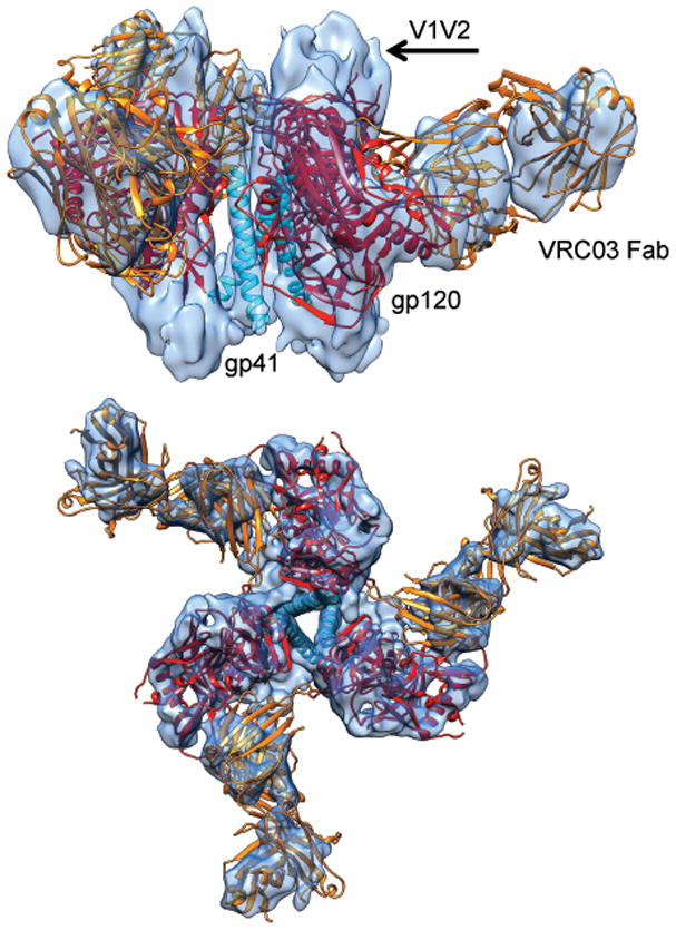
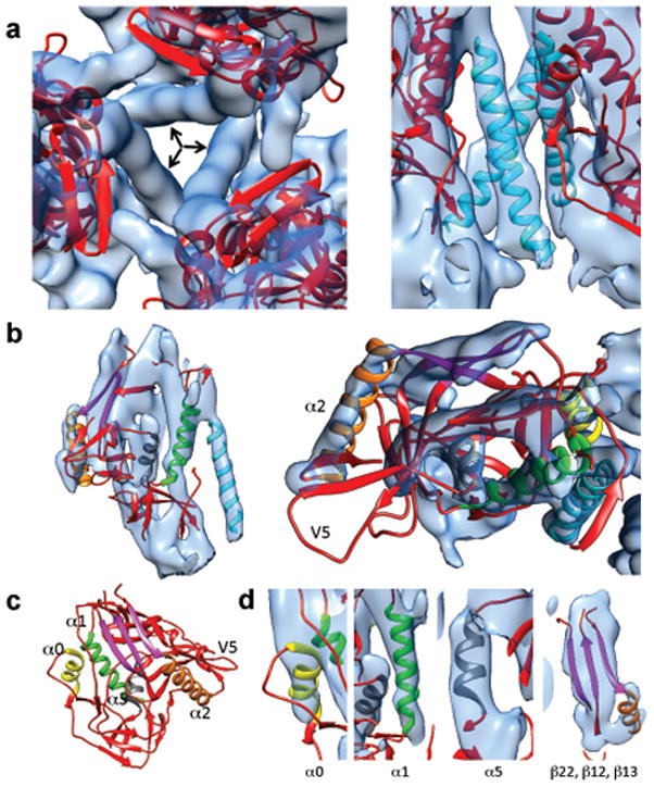
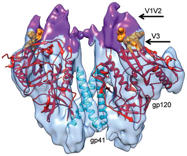
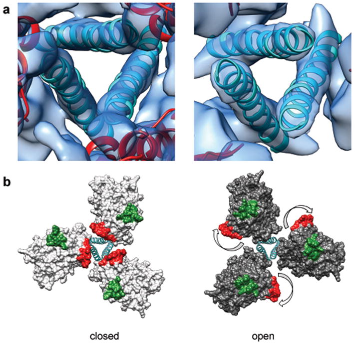
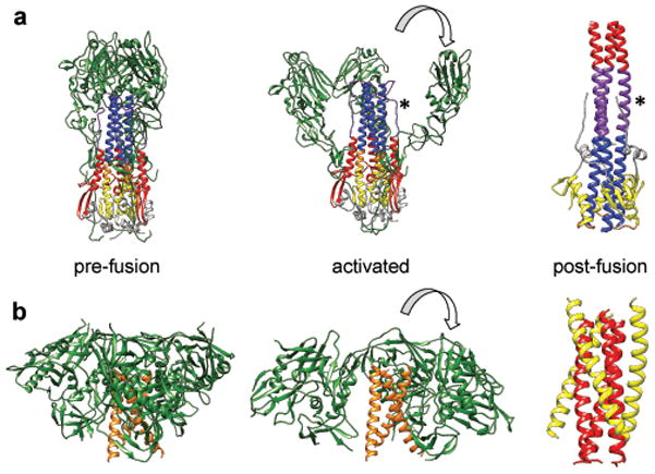
The activation of trimeric HIV-1 envelope glycoprotein (Env) by its binding to the cell-surface receptor CD4 and co-receptors (CCR5 or CXCR4) represents the first of a series of events that lead to fusion between viral and target-cell membranes. Here, we present the cryo-EM structure, at subnanometer resolution (~6 Å at 0.143 FSC), of the 'closed', prefusion state of trimeric HIV-1 Env complexed to the broadly neutralizing antibody VRC03. We show that three gp41 helices at the core of the trimer serve as an anchor around which the rest of Env is reorganized upon activation to the 'open' quaternary conformation. The architecture of trimeric HIV-1 Env in the prefusion state and in the activated intermediate state resembles the corresponding states of influenza hemagglutinin trimers, thus providing direct evidence for the similarity in entry mechanisms used by HIV-1, influenza and related enveloped viruses.


Conflict of interest statement

COMPETING FINANCIAL INTERESTS

The authors declare no competing financial interests.

Figures

Figure 1
Figure 1
Structure of the pre-fusion state of trimeric HIV-1 Env bound to VRC03. Side (top) and top views (bottom) of the density map (from FREALIGN) of soluble HIV-1 Env trimer in complex with Fab fragment of the broadly neutralizing antibody VRC03. Three copies of the X-ray structure (PDB 3SE8) of the complex of the truncated gp120 core (red) with VRC03 Fab (orange), shown as a ribbon representation are fitted into the density map. The three central densities arise from gp41 and are fitted with the long central helix from influenza HA2 (PDB 3HMG)(cyan). Densities that are not interpreted by the ribbon diagrams include the V1V2 loop region, N- and C-terminal ends of gp120 and the portion of gp41 outside of the central rods of density.
Figure 2
Figure 2
Detailed view of gp120 and gp41 structural elements. (a) Close-up of the arrangement of the three gp120 protomers around the three long rods of central density (indicated by arrows) as viewed from the apex of the trimer (left) or the side (right). (b) Close-up of the fit of the gp120 portion of the 3SE8 coordinates into the map shown in side (left) and top views (right). (c) Ribbon diagram of the crystal structure of the gp120 portion of the 3SE8 coordinates highlighting some of the major secondary structural elements α0 (yellow), α1 (green), α2 (orange) and α5 (gray) and a sheet formed from strands β 12, β 13, and β 22 (magenta) in the crystal structure of gp1202. (d) Zoomed-in views of selected regions of the density map illustrating quality of fit of secondary structural elements color-coded as in (c).
Figure 3
Figure 3
Molecular structure of soluble trimeric HIV-1 Env in the closed state. The density for the three VRC03 Fab moieties from the map shown in Figure 1 was computationally removed to provide a view of only the Env component of the complex illustrating the locations of gp120, gp41 and the V1V2 and V3 loops. The density map was fit with three copies of the gp120 core (Chain G of 3SE8), shown in red. Residues 120–204 are not shown and residues at the base of the V3 loop are shown in surface representation and colored orange. The density corresponding to the V1V2 loop is highlighted in purple and the three central gp41 helices (derived from PDB ID 3HMG) are shown in cyan.
Figure 4
Figure 4
Comparison of structures of trimeric Env in the closed, pre-fusion and open, activated conformations. (a) Zoomed-in top views of the closed (left) and open (right) quaternary states derived from structures of trimeric HIV-1 Env in complex with VRC03 and 17b antibodies, respectively. The ribbons representing the central helices are in an identical position in both panels indicating that the location of the central density is approximately the same in closed and open quaternary conformations. (b) Molecular models for the two conformations in (a) show how Env activation results in major rearrangements of gp120 location relative to the central gp41 stalk. The outward rotation of each gp120 protomer repositions the V1V2 loop (base of loop shown in red) from the center to the periphery and alters its position relative to the location of the V3 loop (base of loop shown in green).
Figure 5
Figure 5
Comparison of the structures of influenza hemagglutinin and HIV-1 Env trimers in different states. (a) Structure of hemagglutinin trimer in the pre-fusion state (left panel) displaying the organization of the HA1 trimers (green) around a central stalk formed by HA2 trimers (mixed colors). A model for the initial stages of how the HA1 chains may become repositioned once conformational changes are triggered in hemagglutinin by low pH (middle panel). Structure of post-fusion six-helix bundle state in which the HA2 chain is rearranged (left panel). The HA2 chains are shown in yellow, purple, blue, gray, red and orange to highlight how these regions are rearranged between pre-fusion and post-fusion states. An asterisk is used to help locate the segment of the polypeptide shown in purple that is extended in the middle panel and is helical in the left panel. (b) Structures of trimeric HIV-1 Env in the closed, pre-fusion state described here (left panel), the open, activated state (middle panel) and the post-fusion six-helix bundle state, (right panel). At the present resolution of the maps for the pre-fusion and activated states of HIV-1 Env, we cannot assign different regions of the gp41 polypeptide that correspond to the N-terminal helix region (red) and the C-terminal helix region (yellow).

Similar articles

Cited by

References

    1. Wyatt R, Sodroski J. The HIV-1 envelope glycoproteins: fusogens, antigens, and immunogens. Science. 1998;280:1884–1888. - PubMed
    1. Kwong PD, et al. Structure of an HIV gp120 envelope glycoprotein in complex with the CD4 receptor and a neutralizing human antibody. Nature. 1998;393:648–659. - PMC - PubMed
    1. Huang CC, et al. Structure of a V3-containing HIV-1 gp120 core. Science. 2005;310:1025–1028. - PMC - PubMed
    1. Zhou T, et al. Structural definition of a conserved neutralization epitope on HIV-1 gp120. Nature. 2007;445:732–737. - PMC - PubMed
    1. Pancera M, et al. Structure of HIV-1 gp120 with gp41-interactive region reveals layered envelope architecture and basis of conformational mobility. Proc Natl Acad Sci U S A. 2010;107:1166–1171. - PMC - PubMed

Publication types

Substances

Associated data