. 2014 Jan;111(2):405-14.
doi: 10.1152/jn.00322.2013. Epub 2013 Oct 23.

Modulation of transcallosal inhibition by bilateral activation of agonist and antagonist proximal arm muscles

Affiliations

Modulation of transcallosal inhibition by bilateral activation of agonist and antagonist proximal arm muscles

Monica A Perez et al. J Neurophysiol. 2014 Jan.

Abstract

Transcallosal inhibitory interactions between proximal representations in the primary motor cortex remain poorly understood. In this study, we used transcranial magnetic stimulation to examine the ipsilateral silent period (iSP; a measure of transcallosal inhibition) in the biceps and triceps brachii during unilateral and bilateral isometric voluntary contractions. Healthy volunteers performed 10% of maximal isometric voluntary elbow flexion or extension with one arm while the contralateral arm remained at rest or performed 30% of maximal isometric voluntary elbow flexion or extension. The iSP was measured in the arm performing 10% contractions, and electromyographic (EMG) recordings were comparable across conditions. The iSP onset and duration in the biceps and triceps brachii were comparable. In both muscles, the iSP depth and area were increased during bilateral contractions of homologous agonist muscles (extension-extension and flexion-flexion) compared with a unilateral contraction, whereas during bilateral contractions of nonhomologous antagonist muscles (extension-flexion and flexion-extension), the iSP depth and area were decreased compared with a unilateral contraction, and sometimes facilitation of EMG was seen. This effect was never observed during bilateral activation of homologous muscles. The size of responses evoked by cervicomedullary electrical stimulation in the arm that made 10% contractions remained unchanged across conditions. Thus transcallosal inhibition targeting triceps and biceps brachii is upregulated by voluntary contraction of the contralateral agonist muscle and downregulated by voluntary contraction of the contralateral antagonist muscle. We speculate that these reciprocal task-dependent interactions between bilateral flexor and extensor arm regions of the motor cortex may contribute to coupling between the arms during motor behavior.

Keywords: force; interhemispheric inhibition; primary motor cortex; transcallosal pathways; transcranial magnetic stimulation.


Figures
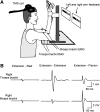
Fig. 1.
Fig. 1.
A: schematic illustration of the experimental setup. Note that feedback of elbow flexion and extension force for the left and right arm (indicated by arrows) was presented on a display in front of the subject. During elbow flexion the visual feedback was provided toward the right side of the screen, and during elbow extension the visual feedback was provided toward the left side of the screen. TMS, transcranial magnetic stimulation. B: electromyography (EMG) of the right biceps and triceps brachii, which were contralateral to the stimulated motor cortex in the elbow extension session. Traces show the average unrectified EMG data in 30 trials. In the left column, recordings are shown during 10% maximal extension on the left (not shown) while the right arm remained at rest (extension-rest). In the middle column, recordings are shown during 10% maximal extension on the left (not shown) and 30% maximal extension with the right arm (extension-extension). In the right column, recordings are shown during 10% maximal extension on the left (not shown) and 30% maximal flexion with the right arm (extension-flexion).
Fig. 2.
Fig. 2.
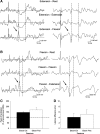
A: raw EMG traces recorded from the triceps brachii muscles in a representative subject during all conditions tested (extension-rest, extension-extension, and extension-flexion) with TMS applied to the ipsilateral motor cortex. Traces show the average rectified (left column) and unrectified (right column) EMG data in 30 trials. The onset and offset of the ipsilateral silent period (iSP) are shown between broken lines. The same time period is also shown on the unrectified data. Notice that the subject shows more inhibition of the EMG during bilateral extension-extension and less inhibition of the EMG during bilateral extension-flexion compared with unilateral elbow extension. B shows individual subject data in all conditions tested; in C, group data (n = 15) show the depth of the iSP in all conditions tested. The abscissa shows the conditions tested, and the ordinate shows the depth of the iSP calculated from the mean EMG activity during the iSP divided by the mean of the prestimulus EMG and subtracted from 100. Note the increase in the iSP during bilateral activation of agonist muscles (extension-extension) and a decrease during bilateral activation of antagonist muscles (extension-flexion) compared with a unilateral contraction. Error bars indicate SE. *P < 0.05. Ext, extension; Flex, flexion.
Fig. 3.
Fig. 3.
A: raw EMG traces recorded from the biceps brachii muscles in a representative subject during all conditions tested (flexion-rest, flexion-flexion, and flexion-extension) with TMS applied to the ipsilateral motor cortex. Traces show the average rectified (left column) and unrectified (right column) EMG data in 30 trials. The onset and offset of the iSP is shown between broken lines. The same time period is also shown on the unrectified data. Notice that the subject shows more inhibition in the EMG during bilateral flexion-flexion and less inhibition in the EMG during flexion-extension compared with unilateral elbow flexion. B shows individual subject data in all conditions tested; in C, group data (n = 15) show the depth of the iSP in all conditions tested. The abscissa shows the conditions tested, and the ordinate shows the depth of the iSP. Similar to what was observed in the triceps, note here the increase in the iSP during bilateral activation of agonist muscles (flexion-flexion) and the decrease during bilateral activation of antagonist muscles (flexion-extension) compared with a unilateral contraction. Error bars indicate SE. *P < 0.05.
Fig. 4.
Fig. 4.
A and B: raw EMG data in 2 representative subjects showing the peak of facilitation (indicated by arrow) preceding the EMG inhibition in the extension-flexion (measured in triceps; A) and flexion-extension conditions (measured in biceps; B). The peak is not present in the other conditions tested. C: the abscissa shows the sessions tested (elbow extension, n = 5; elbow flexion, n = 4) and the ordinate shows the peak of facilitation (expressed as a percentage of the prestimulus EMG). D: the abscissa shows the sessions tested (elbow extension, n = 5; elbow flexion, n = 4) and the ordinate shows the latency of the facilitation (beginning defined as 2 SD above the mean rectified EMG for a period of 90 ms before the TMS) minus the latency of the iSP. Note that the magnitude and the latency of the peak of facilitation observed during bilateral activation of antagonist muscles were similar in both sessions. Error bars indicate SE. *P < 0.05.
Fig. 5.
Fig. 5.
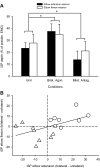
A: a comparison of the iSP in the triceps and biceps measured in the same subjects (n = 8) in all conditions tested. The abscissa shows the conditions tested (Unil, unilateral; Bilat. Agon., bilateral activation of agonist muscles; Bilat. Antag., bilateral activation of antagonist muscles) during the elbow extension (filled bars) and flexion sessions (open bars). The ordinate shows the depth of the iSP. Note the increase in the iSP during bilateral activation of agonist muscles and a decreased during bilateral activation of antagonist muscles compared with a unilateral contraction regardless of the elbow session tested. Error bars indicate SE. *P < 0.05. B: a correlation analysis between flexion and extension sessions of the changes in the iSP depth during bilateral (△, agonist; ○, antagonist) compared with unilateral contractions (a negative number indicates more inhibition and a positive number indicates less inhibition during the unilateral compared with bilateral condition). Each subject is represented by 2 points: one for bilateral agonist and one for bilateral antagonist contractions. Note that subjects who showed more pronounced inhibition in the elbow extension session also showed a similar trend in the elbow flexion session.
Fig. 6.
Fig. 6.
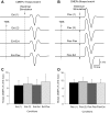
A and B: raw EMG data in 2 representative subjects showing motor evoked responses to electrical cervicomedullary stimulation (cervicomedullary motor evoked potentials, CMEPs) recorded from the triceps (A) and biceps brachii (B) in all conditions tested in the elbow extension [Ext (1), extension-rest, CMEPs alone; Ext (2), extension-rest, CMEPs preceded by TMS; Ext-Ext, extension-extension, CMEPs preceded by TMS; Ext-Flex, extension-flexion, CMEPs preceded by TMS] and flexion sessions [Flex (1), flexion-rest, CMEPs alone; Flex (2), flexion-rest, CMEPs preceded by TMS; Flex-Flex, flexion-flexion, CMEPs preceded by TMS; flex-Ext, flexion-extension, CMEPs preceded by TMS]. Ten traces were average in each set. Because the stimulation artifact in the triceps brachii is small, the time of stimulation is indicated by dashed lines. C and D: in the graphs, the abscissa shows the conditions tested and the ordinate shows the size of CMEPs as a percentage of the maximal M wave (Mmax) in the triceps (C) and biceps brachii (D). Note that the size of CMEPs in triceps and biceps remained similar across conditions in both sessions. Error bars indicate SE. *P < 0.05.

Similar articles

Cited by

References

    1. Aimonetti JM, Nielsen JB. Changes in intracortical excitability induced by stimulation of wrist afferents in man. J Physiol 534: 891–902, 2001 - PMC - PubMed
    1. Bäumer T, Bock F, Koch G, Lange R, Rothwell JC, Siebner HR, Münchau A. Magnetic stimulation of human premotor or motor cortex produces interhemispheric facilitation through distinct pathways. J Physiol 572: 857–868, 2006 - PMC - PubMed
    1. Bäumer T, Dammann E, Bock F, Klöppel S, Siebner HR, Münchau A. Laterality of interhemispheric inhibition depends on handedness. Exp Brain Res 180:195–203, 2007 - PubMed
    1. Bawa P, Hamm JD, Dhillon P, Gross PA. Bilateral responses of upper limb muscles to transcranial magnetic stimulation in human subjects. Exp Brain Res 158: 385–390, 2004 - PubMed
    1. Bestmann S, Swayne O, Blankenburg F, Ruff CC, Haggard P, Weiskopf N, Josephs O, Driver J, Rothwell JC, Ward NS. Dorsal premotor cortex exerts state-dependent causal influences on activity in contralateral primary motor and dorsal premotor cortex. Cereb Cortex 18: 1281–1291, 2008 - PMC - PubMed

Publication types

LinkOut - more resources