Review
. 2014 May;306(4):313-330.
doi: 10.1007/s00403-013-1416-8. Epub 2013 Oct 24.

NADPH oxidase enzymes in skin fibrosis: molecular targets and therapeutic agents

Affiliations
Review

NADPH oxidase enzymes in skin fibrosis: molecular targets and therapeutic agents

Olubukola Babalola et al. Arch Dermatol Res. 2014 May.

Abstract

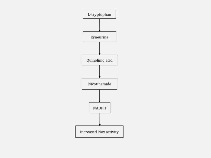
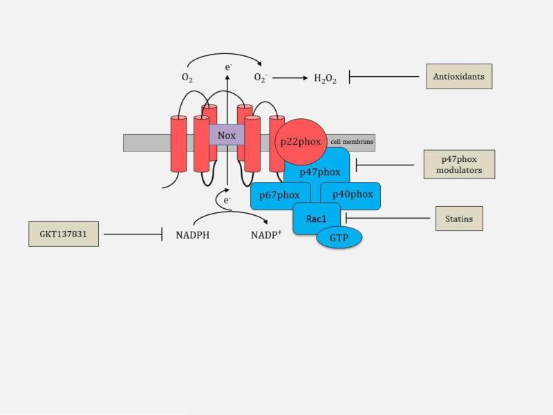
Fibrosis is characterized by the excessive deposition of extracellular matrix components eventually resulting in organ dysfunction and failure. In dermatology, fibrosis is the hallmark component of many skin diseases, including systemic sclerosis, graft-versus-host disease, hypertrophic scars, keloids, nephrogenic systemic fibrosis, porphyria cutanea tarda, restrictive dermopathy and other conditions. Fibrotic skin disorders may be debilitating and impair quality of life. There are few FDA-approved anti-fibrotic drugs; thus, research in this area is crucial in addressing this deficiency. Recent investigations elucidating the pathogenesis of skin fibrosis have implicated endogenous reactive oxygen species produced by the multicomponent nicotinamide adenine dinucleotide phosphate (NADPH) oxidase (Nox) enzyme complex. In this review, we discuss Nox enzymes and their role in skin fibrosis. An overview of the Nox enzyme family is presented and their role in the pathogenesis of skin fibrosis is discussed. The mechanisms by which Nox enzymes influence specific fibrotic skin disorders are also reviewed. Finally, we describe the therapeutic approaches to ameliorate skin fibrosis by directly targeting Nox enzymes with the use of statins, p47phox subunit modulators, or GKT137831, a competitive inhibitor of Nox enzymes. Nox enzymes can also be targeted indirectly via scavenging ROS with antioxidants. We believe that Nox modulators are worthy of further investigation and have the potential to transform the management of skin fibrosis by dermatologists.


Figures

Fig. 1
Fig. 1
Overview of fibrosis. Fibrosis is triggered by an insult such as damage to the endothelial cells in the vasculature. These damaged endothelial cells secrete cytokines resulting in the recruitment of immune cells. Immune cells produce transforming growth factor (TGF)-beta1, connective tissue growth factor (CTGF), and platelet-derived growth factor (PDGF) leading to fibroblast activation, proliferation and differentiation into myofibroblasts. The downstream effect is excessive extracellular matrix (ECM) synthesis and deposition by fibroblasts and myofibroblasts, a hallmark feature of fibrosis. IL, interleukin; TNF, tumor necrosis factor.
Fig. 2
Fig. 2
Structural components and molecular organization of the Nox family. Part a shows the molecular structure of Nox2. Rac1 is not membrane anchored and it binds to GTP. Part b shows the enzymatic reaction catalyzed by Nox enzymes. Nox1, Nox2 and Nox5 generate the superoxide anion (O2); Nox4, Duox1 and Duox2 further dismutase O2 to hydrogen peroxide (H2O2) as their final product. Part c shows the molecular structure of Nox1; Nox3 has a similar structure to Nox1. Part d shows the molecular structure of Nox4. Parts e-f show the molecular structures of Nox5, Duox1 and Duox2 - these three enzymes have the similar structures with intracellular calcium (Ca) binding N-terminal cytoplasmic EF-hand domain regions e, electron; FAD, flavin adenine dinucleotide; O2, molecular oxygen; NOXA1, Nox activator 1; NOXO1, Nox organizer 1; POLDIP2, DNA polymerase-delta-interacting protein; Ca, calcium.
Fig. 3
Fig. 3
Flowchart illustrating the literature search strategy and results.
Fig. 4
Fig. 4
Mechanistic Involvement of Nox in skin fibrosis. Nox enzymes influence several pathways that result in skin fibrosis. In toxic oil syndrome, 5-VPTA stimulates PKC to activate Nox. Nox enzymes produce ROS that inactivate the proapoptic caspases promoting survival of fibroblasts, the key cell involved in matrix deposition. ROS also inactivates protein tyrosine phosphatases to promote the activities of the tyrosine kinases, JNK, MAPK, JAK, c-Src, and ERK (EGCG modulates the tyrosine kinases). Inhibition of the protein tyrosine phosphatase-1B by ROS increases the activity of the tyrosine kinase, PDGFR. PDGFR's interaction with PDGF further upregulates ERK. Autoantibodies (Ab), identified in scleroderma, stimulate the PDGFR and its downstream pathway, ERK. PDGFR induces Nox1 and Nox2 and autoantibodies enhance this induction. All these tyrosine kinases enhance the transcription of genes involved in skin fibrosis, including TGF-beta, CTGF, PDGF and ECM proteins. ROS promotes the activation of MMPs in the process termed cysteine switch. ROS also upregulates expression of the chemokine ligand-2 (CCL-2) implicated in scleroderma and GVHD; statins antagonize this upregulation. ROS promotes telomere shortening, a process implicated in keloids and restrictive dermopathy. 5-VPTA, N-(5-vinyl-1,3-thiazolidin-2-ylidene) phenylamine; PKC, protein kinase C; PDGF, platelet-derived growth factor; PDGFR, PDGF receptor; EGCG, epigallocatechin-3-gallate; Zn, zinc; C, cysteine.
Fig. 5
Fig. 5
Metabolism of L-tryptophan to NADPH in eosinophilia-myalgia syndrome (EMS). Increased ingestion of L-tryptophan in EMS results in increased NADPH concentration with an associated increase in the activity of Nox enzymes.
Fig. 6
Fig. 6
Nox-associated therapeutic agents in skin fibrosis. Therapeutic agents can be directed at modulating the p47phox subunit, inhibiting the migration of Rac1 to the cell membrane (statins), competitively inhibiting Nox with molecules mimicking NADPH (GKT137831), or scavenging reactive oxygen species, O2 and H2O2 (antioxidants).

References

    1. Abdullah A, Blakeney P, Hunt R, Broemeling L, Phillips L, Herndon DN, Robson MC. Visible scars and self-esteem in pediatric patients with burns. The Journal of burn care & rehabilitation. 1994;15(2):164–168. - PubMed
    1. Akasaki T, Ohya Y, Kuroda J, Eto K, Abe I, Sumimoto H, Iida M. Increased expression of gp91phox homologues of NAD(P)H oxidase in the aortic media during chronic hypertension: involvement of the renin-angiotensin system. Hypertension research : official journal of the Japanese Society of Hypertension. 2006;29(10):813–820. doi:10.1291/hypres.29.813. - PubMed
    1. Alexandre J, Hu Y, Lu W, Pelicano H, Huang P. Novel action of paclitaxel against cancer cells: bystander effect mediated by reactive oxygen species. Cancer research. 2007;67(8):3512–3517. doi:10.1158/0008-5472.CAN-06-3914. - PubMed
    1. Altenhofer S, Kleikers PW, Radermacher KA, Scheurer P, Rob Hermans JJ, Schiffers P, Ho H, Wingler K, Schmidt HH. The NOX toolbox: validating the role of NADPH oxidases in physiology and disease. Cellular and molecular life sciences : CMLS. 2012;69(14):2327–2343. doi:10.1007/s00018-012-1010-9. - PMC - PubMed
    1. Aoyama T, Paik YH, Watanabe S, Laleu B, Gaggini F, Fioraso-Cartier L, Molango S, Heitz F, Merlot C, Szyndralewiez C, Page P, Brenner DA. Nicotinamide adenine dinucleotide phosphate oxidase in experimental liver fibrosis: GKT137831 as a novel potential therapeutic agent. Hepatology. 2012;56(6):2316–2327. doi:10.1002/hep.25938. - PMC - PubMed

Publication types