Review
. 2014 Jan;23(1):1-22.
doi: 10.1002/pro.2385. Epub 2013 Nov 18.

Cholesterol as a co-solvent and a ligand for membrane proteins

Affiliations
Review

Cholesterol as a co-solvent and a ligand for membrane proteins

Yuanli Song et al. Protein Sci. 2014 Jan.

Abstract

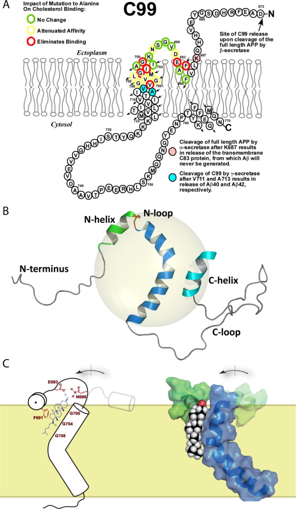
As of mid 2013 a Medline search on "cholesterol" yielded over 200,000 hits, reflecting the prominence of this lipid in numerous aspects of animal cell biology and physiology under conditions of health and disease. Aberrations in cholesterol homeostasis underlie both a number of rare genetic disorders and contribute to common sporadic and complex disorders including heart disease, stroke, type II diabetes, and Alzheimer's disease. The corresponding author of this review and his lab stumbled only recently into the sprawling area of cholesterol research when they discovered that the amyloid precursor protein (APP) binds cholesterol, a topic covered by the Hans Neurath Award lecture at the 2013 Protein Society Meeting. Here, we first provide a brief overview of cholesterol-protein interactions and then offer our perspective on how and why binding of cholesterol to APP and its C99 domain (β-CTF) promotes the amyloidogenic pathway, which is closely related to the etiology of Alzheimer's disease.

Keywords: Alzheimer's disease; C99; CRAC; GPCRs; amyloid precursor protein; cholesterol; integral membrane proteins; receptor; β-CTF.


Figures

Figure 1
Figure 1
Biosynthesis of sterols in eukaryotes and sterol surrogates in prokaryotes share all steps up to the intermediate, squalene.
Figure 2
Figure 2
Structure of cholesterol. A) Chemical structure of cholesterol (IUPAC numbering system). B) Space-filling and stick representations of cholesterol. C) Superposition of three cholesterol molecules from different protein crystal structures demonstrates the flexibility of the tail.
Figure 3
Figure 3
Structures of representative lipids: 1-palmitoyl-2-oleoyl-sn-glycero-3-phosphocholine (POPC), 1-palmitoyl-2-oleoyl-sn-glycero-3-phosphoglycerol (POPG), 1-palmitoyl-2-oleoyl-sn-glycero-3-phosphserine (POPS), and 1-palmitoyl-2-oleoyl-sn-glycero-3-phosphoethanolamine (POPE), sphingomyelin, and cholesterol. The atoms in the membrane interface region colored in red are possible hydrogen bond acceptors. Atoms in blue are possible hydrogen bond donors.
Figure 4
Figure 4
Lipids and water as solvents. A) Cartoon model of a soluble protein in a bath of water. Bulk water molecules are shown in grey; shell water molecules are shown in yellow; a bound water molecule is shown in red. B) Cartoon model of a membrane protein embedded in a membrane bilayer. Bulk lipids are shown in grey; annular lipids are shown in yellow; a bound (non-annular) lipid is shown in red.
Figure 5
Figure 5
General modes of cholesterol binding to proteins. A) Cholesterol bound to a cavity in the yeast Osh4 protein (PDB ID: 1ZHY90). B) Cholesterol bound to the surface of a μ-type opioid receptor (PDB ID: 4DKL101).
Figure 6
Figure 6
Amino acid composition of structurally-defined cholesterol binding sties. A) Occurrence of amino acids in all proteins (cyan bar) versus in the 19 cholesterol-associated proteins of Table I (white bar). B–G) Occurrence in the PDB of amino acids within 5 Å of different moieties within cholesterol. The y-axis % occurrence (left) and absolute number of observations (right) reflect the number of times that at least one atom from that amino acid (including backbone atoms) is within 5 Å of at least one atom of the indicated substituent moiety of cholesterol.
Figure 7
Figure 7
Cholesterol usually resides on the TM surface of integral membrane proteins. α) Cholesterol binds to TM helix I of the 5-hydroxytryptamine receptor 2B (PDBID: 4IB4100). B) Cholesterol binds to the groove formed by TM helices VI and VII of the μ-type opioid receptor (PDBID: 4DKL101). C) The tail of cholesterol 1 in a Na+, K+-ATPase (PDBID: 4HYT94) fits the crevice between TM helix 7 and 10. D) The tail of cholesterol 2 in a Na+, K+-ATPase (PDBID: 4HYT94) fits the gap between TM helix 8 and 10. The C5=C6 double bond in ring B of cholesterol is shown in magenta.
Figure 8
Figure 8
Cholesterol resides on the surface of 5-hydroxytryptamine receptor 2B (PDBID: 4IB4100) with its β-face contacting the surface of the protein (atoms shown in yellow) and its α-face contacting acyl chains of monoolein (cyan) and the C16 chain of covalently-linked palmitate (atoms shown in white).
Figure 9
Figure 9
Examples of hydrogen bonds formed between the head group of bound cholesterol and proteins. A) Cholesterol forms a hydrogen bond with the side chain of Gln65 in the β2-adrenergic receptor (PDB ID: 3PDS98). B) Cholesterol forms a hydrogen bond with the backbone amide Tyr394 in the 5-hydroxytryptamine receptor 2B (PDBID: 4IB4 100). C) Cholesterol forms a hydrogen bond network with Pro309 and involves a water molecule (shown as a red ball) in the μ-type opioid receptor (PDBID: 4DKL101). D) Two cholesterols form a hydrogen bond network with Gln65, Tyr70, and Arg151 through a water molecule (shown as a red ball) in the β2 adrenergic receptor (PDBID: 3NYA96). E) Cholesterol forms a hydrogen network with Glu30, Asn41, and Gln79 in the Niemann-Pick C1 protein (PDBID: 3GKI89) mediated by a water molecule (shown as a red ball). F) Cholesterol forms a hydrogen bond network with Tyr61, Asn210, and Gln377 in human CYP11A1 (PDBID: 3N9Y91) through multiple water molecules (shown as red balls).
Figure 10
Figure 10
Examples of an aromatic residue (shown in magenta) interacting with the sterol ring of cholesterol. A) Phe33 interacts the α face of the sterol ring in a Na+, K+-ATPase (PDBID: 4HYT, cholesterol 1 94). B) Trp981 interacts the β-face of cholesterol in a Na+, K+-ATPase (PDBID: 4HYT, cholesterol 2 94). C) Phe255 interacts the α face of the sterol ring in the adenosine receptor A2α (PDB ID: 4EIY102). D) Trp158 interacts the α-face of cholesterol in the β2-adrenergic receptor (PDB ID: 2RH1). All carbon atoms in cholesterol and aromatic residues are displayed with spheres with a radius of 1.7 Å. E) Cholesterol sandwich packing configuration with Phe255 in the middle and cholesterols on each side in the adenosine receptor A2α (PDBID: 4EIY102). The C5=C6 double bonds (shown in orange) are parallel to the face of the aromatic ring. F) Aromatic sandwich packing configuration with a cholesterol in the middle and Tyr299 and Phe313 on each side in the μ-type opioid receptor (PDBID: 4DKL101). The C5=C6 double bond (shown in orange) is parallel to the faces of the aromatic rings. G) Interaction between the Phe203 ring of the Niemann-Pick C1 protein and the cholesterol double bond (PDBID: 3GKI89). H) Interaction of F82 of with the C5=C6 group of CYP11A1 (a cytochrome p450, PDBID: 3N9Y91).
Figure 11
Figure 11
Cholesterol-proximal CRAC motifs in the Osh4 protein. A) Three CRAC motifs are highlighted in the Osh4 protein (PDB ID: 1ZHY90), with one motif (shown in blue) far from the cholesterol and the other two (shown in red) close to the cholesterol. Side chains for key residues of the closest CRAC motifs are shown. B) The cholesterol consensus motif (CCM) in the β2-adrenergic acceptor (PDB ID: 3D4S99). Cholesterol 1 binds to the CCM primarily in contact with the fourth transmembrane helix. Side chains for key residues in the CCM are shown. I1544.46 and W1584.50 are also shown in space-filling mode and interact with cholesterol 1. R1514.43 and Y702.41 may form a hydrogen bonding network, as shown with black dotted lines. The two superscript numbers for each residue reflect the Ballesteros-Weinstein numbering scheme for GPCR sites. The first number indicates the transmembrane helix, while the second is relative to the most conserved residue in that helix, which is designated position 50. For example, residue 4.46I is the fourth residue before the most conserved residue in helix IV (4.50W in this particular case).
Figure 12
Figure 12
Structure of C99 and formation of a complex with cholesterol. A) The topology of C99. C99 is composed of the C-terminal 99 residues domain of the amyloid precursor protein (residues 672–770), and contains the cleavage site for α-secretase (following K687 as shown in pink) and cleavage sites for γ-secretase (following V711 and A713, as shown in cyan). Alanine-scanning of C99 residues 690 to 710 revealed which residues are critical for cholesterol binding, as indicated. This panel was adapted from Reference 115, with permission from American Chemical Society. B) Backbone structure of C99, as determined by NMR for the protein in LMPG micelles (represented with the gray sphere). This panel was adapted from Reference 116, with permission from American Association for the Advancement of Science. C) Rough model for the proposed C99-cholesterol complex and related conformational change in C99. This panel was adapted from Reference 116, with permission from American Association for the Advancement of Science (left side) and from Reference 115, with permission from American Chemical Society (right side).

References

    1. Evans WH, Hardison WG. Phospholipid, cholesterol, polypeptide and glycoprotein composition of hepatic endosome subfractions. Biochem J. 1985;232:33–36. - PMC - PubMed
    1. Zambrano F, Fleischer S, Fleischer B. Lipid composition of the Golgi apparatus of rat kidney and liver in comparison with other subcellular organelles. Biochim Biophys Acta. 1975;380:357–369. - PubMed
    1. Mitra K, Ubarretxena-Belandia I, Taguchi T, Warren G, Engelman DM. Modulation of the bilayer thickness of exocytic pathway membranes by membrane proteins rather than cholesterol. Proc Natl Acad Sci USA. 2004;101:4083–4088. - PMC - PubMed
    1. Andreyev AY, Fahy E, Guan Z, Kelly S, Li X, McDonald JG, Milne S, Myers D, Park H, Ryan A, Thompson BM, Wang E, Zhao Y, Brown HA, Merrill AH, Raetz CR, Russell DW, Subramaniam S, Dennis EA. Subcellular organelle lipidomics in TLR-4-activated macrophages. J Lipid Res. 2010;51:2785–2797. - PMC - PubMed
    1. Wood WG, Cornwell M, Williamson LS. High performance thin-layer chromatography and densitometry of synaptic plasma membrane lipids. J Lipid Res. 1989;30:775–779. - PubMed

Publication types