

Contribution of mesenchymal proliferation in tooth root morphogenesis
- PMID: 24155265
- PMCID: PMC3872847
- DOI: 10.1177/0022034513511247
Contribution of mesenchymal proliferation in tooth root morphogenesis
Abstract
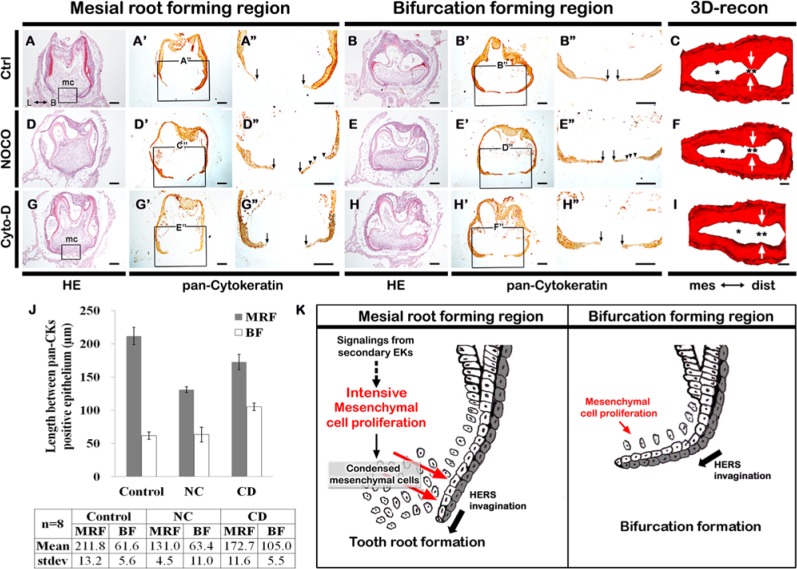
In mouse tooth development, the roots of the first lower molar develop after crown formation to form 2 cylindrical roots by post-natal day 5. This study compared the morphogenesis and cellular events of the mesial-root-forming (MRF) and bifurcation-forming (BF) regions, located in the mesial and center of the first lower molar, to better define the developmental mechanisms involved in multi-rooted tooth formation. We found that the mesenchyme in the MRF showed relatively higher proliferation than the bifurcation region. This suggested that spatially regulated mesenchymal proliferation is required for creating cylindrical root structure. The mechanism may involve the mesenchyme forming a physical barrier to epithelial invagination of Hertwig's epithelial root sheath. To test these ideas, we cultured roots in the presence of pharmacological inhibitors of microtubule and actin polymerization, nocodazole and cytochalasin-D. Cytochalasin D also inhibits proliferation in epithelium and mesenchyme. Both drugs resulted in altered morphological changes in the tooth root structures. In particular, the nocodazole- and cytochalasin-D-treated specimens showed a loss of root diameter and formation of a single-root, respectively. Immunolocalization and three-dimensional reconstruction results confirmed these mesenchymal cellular events, with higher proliferation in MRF in multi-rooted tooth formation.
Keywords: cell proliferation; histochemistry; molar; odontogenesis; tooth development; tooth root.
Conflict of interest statement
The authors declare no potential conflicts of interest with respect to the authorship and/or publication of this article.
Figures


References
-
- Fujiwara N, Tabata MJ, Endoh M, Ishizeki K, Nawa T. (2005). Insulin-like growth factor-I stimulates cell proliferation in the outer layer of Hertwig’s epithelial root sheath and elongation of the tooth root in mouse molars in vitro. Cell Tissue Res 320:69-75 - PubMed
-
- Hosoya A, Kim JY, Cho SW, Jung HS. (2008). BMP4 signaling regulates formation of Hertwig’s epithelial root sheath during tooth root development. Cell Tissue Res 333:503-509 - PubMed
-
- Ishikawa Y, Ida-Yonemochi H, Nakakura-Ohshima K, Ohshima H. (2012). The relationship between cell proliferation and differentiation and mapping of putative dental pulp stem/progenitor cells during mouse molar development by chasing BrdU-labeling. Cell Tissue Res 348:95-107 - PubMed
-
- Kim JY, Cho SW, Song WC, Lee MJ, Cai J, Ohk SH, et al. (2005). Formation of spacing pattern and morphogenesis of chick feather buds is regulated by cytoskeletal structures. Differentiation 73:240-248 - PubMed
Publication types
MeSH terms
Substances
LinkOut - more resources
Full Text Sources
Other Literature Sources
Miscellaneous
