. 2013 Oct 23;33(43):16865-73.
doi: 10.1523/JNEUROSCI.2449-13.2013.

The infralimbic cortex bidirectionally modulates mesolimbic dopamine neuron activity via distinct neural pathways

Affiliations

The infralimbic cortex bidirectionally modulates mesolimbic dopamine neuron activity via distinct neural pathways

Mary H Patton et al. J Neurosci. .

Abstract

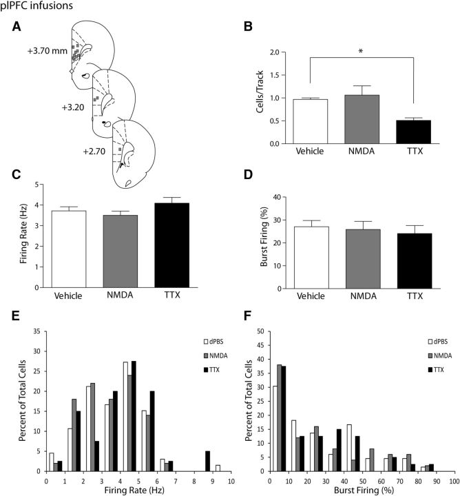
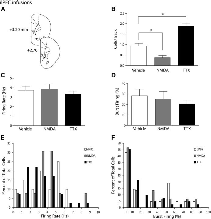
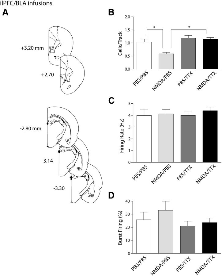
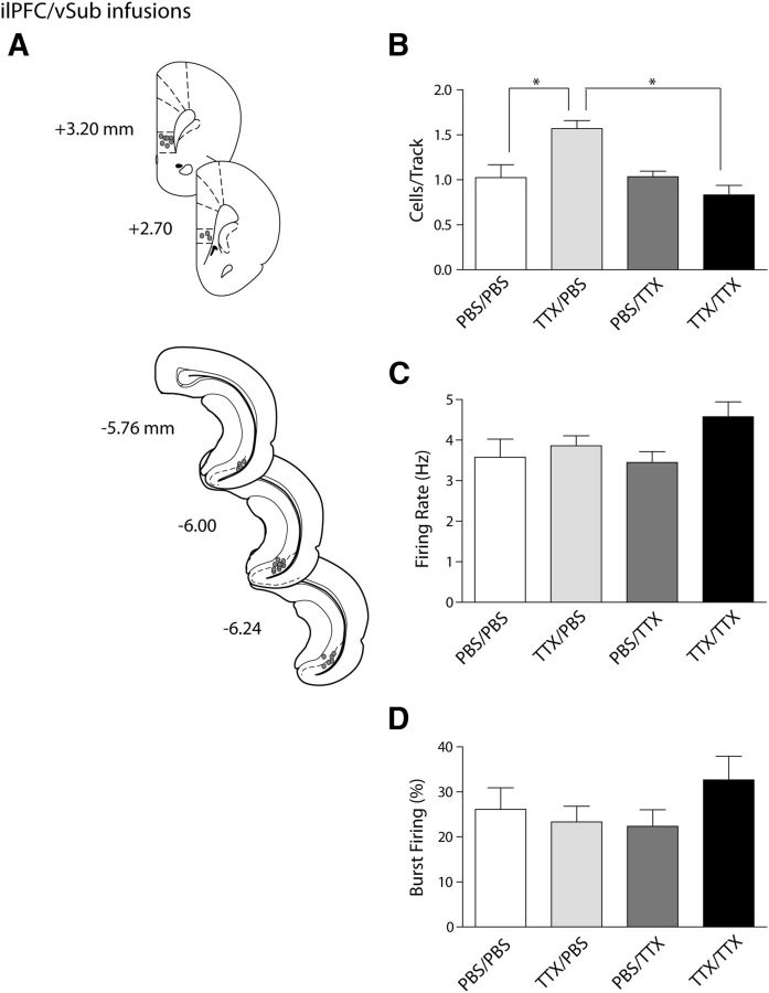
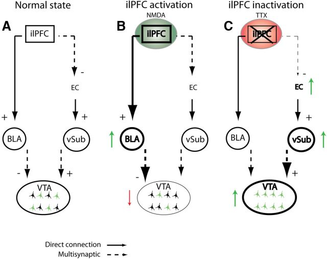
The ventral tegmental area (VTA) has been implicated in a number of psychiatric disorders, including schizophrenia, depression, and bipolar disorder. One major regulator of the mesolimbic dopaminergic system is the medial prefrontal cortex (mPFC), which makes direct and indirect connections to the hippocampus and amygdala, as well as directly to the VTA. The mPFC is comprised of two subregions: the infralimbic and prelimbic cortices (ilPFC and plPFC). However, the specific roles of these subregions in regulating VTA dopamine activity have remained unclear. In this study, we aim to clarify this role and to examine the divergent neuranatomical circuits by which the mPFC regulates VTA activity. Using in vivo extracellular recordings in rats, we tested the effects of pharmacological activation (with NMDA) and inactivation (with TTX) of the ilPFC and plPFC on dopamine neuron activity, and tested the roles of the ventral subiculum (vSub) and basolateral amygdala in this process. We found that the ilPFC exerts a bidirectional control of VTA dopamine neurons, which are differentially modulated through the vSub and the basolateral amygdala. Specifically, activation or inactivation of the ilPFC attenuated or activated dopamine neuron population activity, respectively. Furthermore, dopamine activation depended on the ventral hippocampus and inactivation on the amygdala. In contrast, only inactivation of the plPFC altered dopamine neuron activity. These data indicate that the mPFC has the ability to uniquely fine-tune dopaminergic activity in the VTA. Furthermore, the data presented here suggest that the ilPFC may have a role in the pathophysiology of psychiatric disorders.


Figures

Figure 1.
Figure 1.
Inactivation of the plPFC produces a selective attenuation of VTA DA neuron population activity. A, Representation of histological placements of infusion cannulae into the plPFC (squares, ∼50% shown). B, Activating the plPFC with NMDA did not change the number of spontaneously active dopamine neurons firing in the VTA (expressed as cells/track, gray bar) compared with infusion of vehicle (white bar). Inactivating the plPFC with TTX resulted in a significant decrease in the number of spontaneously active dopamine cells in the VTA (black bar). C, D, The firing rate of spontaneously active dopamine cells and the percentage of cells firing in bursts were not affected by infusion of vehicle (white bar), NMDA (gray bar), or TTX (black bar) into the plPFC. Distributions of firing rate (E) and the percentage of burst firing (F) were not affected significantly by infusions of NMDA or TTX (Kolgomorov–Smirnoff test). *p < 0.05 (one-way ANOVA, Holm-Sidak post hoc test). n = 7–10 rats/group; n = 40–66 neurons/group. Data are represented as mean ± SEM.
Figure 2.
Figure 2.
Activation or inactivation of the ilPFC selectively decreases or increases VTA DA neuron population activity, respectively. A, Representation of histological placements of infusion cannulae in the ilPFC (circles, ∼50% shown). B, Activating the ilPFC with NMDA resulted in a significant decrease in the number of spontaneously active DA neurons in the VTA (cells/track, gray bar), whereas inactivating the ilPFC with TTX yielded a significant increase in the number of spontaneously active DA neurons in the VTA (black bar), compared with vehicle injections (white bar). C, D, Infusion of vehicle (white bars), NMDA (gray bars), or TTX (black bars) led to no changes in the firing rate or percentage of spikes in bursts in dopaminergic cells in the VTA. Distributions of firing rate (E) and the percentage of burst firing (F) were not affected significantly by infusions of NMDA or TTX (Kolgomorov–Smirnoff test). *p < 0.05 (one-way ANOVA, Holm-Sidak post hoc test). n = 4 or 5 rats/group; n = 15–42 neurons/group. Data are represented as mean ± SEM.
Figure 3.
Figure 3.
The ilPFC activation-induced decrease in VTA DA neuron population activity is mediated via the BLA. A, Histological placement of infusion cannulae into the ilPFC and BLA (circles, ∼50% of ilPFC placements [top] shown for clarity and 100% of BLA placements [bottom] shown). B, Dual infusions of NMDA into the ilPFC and TTX into the BLA (black bar) prevented the decrease in number of spontaneously active dopamine cells in the VTA that occurred with activation of the ilPFC alone (cells/track, light gray bar). Inactivating the BLA alone did not yield any changes in dopamine population activity compared with vehicle infusions (dark gray bar and white bar, respectively). C, D, The firing rate and percentage of spikes occurring in bursts were not affected by any of the infusions. *p < 0.05 (two-way ANOVA, Bonferroni post hoc test). n = 5 rats/group; n = 18–41 neurons/group. Data are represented as mean ± SEM.
Figure 4.
Figure 4.
The ilPFC inactivation-induced increase in VTA DA neuron population activity is mediated via the vSub. A, Histological placement of infusion cannulae in the ilPFC and vSub (circles, ∼50% of ilPFC placements [top] shown for clarity, and 100% of vSub placements [bottom] shown). B, Inactivating both the ilPFC and the vSub with TTX (black bar) prevented the hyperactivity of dopamine population activity in the VTA that occurred with inactivation of the ilPFC alone (cells/track, light gray bar). Inactivating the vSub had no effect on the overall population activity (dark gray bar) compared with vehicle infusions (white bar). C, D, The firing rate and percentage of spikes occurring in bursts were not affected by any of the infusion treatments. *p < 0.05 (two-way ANOVA, Bonferroni post hoc test). n = 5 or 6 rats/group; n = 29–49 neurons/group. Data are represented as mean ± SEM.
Figure 5.
Figure 5.
Diagram representing two proposed pathways through which the ilPFC could act to modulate the activity of DA neurons in the VTA. A, Normal pathway without any manipulation to any regions shows the normal population activity in the VTA (green colored neurons indicate spontaneously active DA cells). B, Activation of the ilPFC with NMDA (green circle) leads to a decrease in population activity (red downward arrow) in the VTA via activation of the BLA pathway (thicker lines and green upward arrow). Dashed lines from the BLA to the VTA indicate that this pathway has not yet been delineated. C, Inactivation of the ilPFC with TTX (red circle) leads to an increase in population activity in the VTA (green upward arrow) potentially through attenuation of an indirect inhibitory pathway from the ilPFC to the entorhinal cortex (EC), leading to an increase in vSub firing and VTA population activity (green upward arrows). The dashed line from the vSub to the VTA indicates a multisynaptic pathway that includes the nucleus accumbens and ventral pallidum (Floresco et al., 2003).

References

    1. Amat J, Baratta MV, Paul E, Bland ST, Watkins LR, Maier SF. Medial prefrontal cortex determines how stressor controllability affects behavior and dorsal raphe nucleus. Nat Neurosci. 2005;8:365–371. doi: 10.1038/nn1399. - DOI - PubMed
    1. American Psychiatric Association. Diagnostic and statistical manual of mental disorders, Ed 4 (Text Revision) American Psychiatric Association: Washington, DC; 2000.
    1. Bora E, Fornito A, Yücel A, Pantelis C. Voxelwise meta-analysis of gray matter abnormalities in bipolar disorder. Biol Psychiatry. 2010;67:1097–1105. doi: 10.1016/j.biopsych.2010.01.020. - DOI - PubMed
    1. Brooks JO, 3rd, Bonner JC, Rosen AC, Wang PW, Hoblyn JC, Hill SJ, Ketter TA. Dorsolateral and dorsomedial prefrontal gray matter density changes associated with bipolar depression. Psychiatry Res. 2009;172:200–204. doi: 10.1016/j.pscychresns.2008.06.007. - DOI - PMC - PubMed
    1. Chang CH, Grace AA. Amygdala β-noradrenergic receptors modulate delayed downregulation of dopamine activity following restraint. J Neurosci. 2013;33:1441–1450. doi: 10.1523/JNEUROSCI.2420-12.2013. - DOI - PMC - PubMed

Publication types

MeSH terms

LinkOut - more resources