. 2013 Oct 23;33(43):16930-44.
doi: 10.1523/JNEUROSCI.3513-12.2013.

Regulator of calcineurin 1 modulates expression of innate anxiety and anxiogenic responses to selective serotonin reuptake inhibitor treatment

Affiliations

Regulator of calcineurin 1 modulates expression of innate anxiety and anxiogenic responses to selective serotonin reuptake inhibitor treatment

Charles A Hoeffer et al. J Neurosci. .

Abstract

Regulator of calcineurin 1 (RCAN1) controls the activity of calcium/calmodulin-dependent phosphatase calcineurin (CaN), which has been implicated in human anxiety disorders. Previously, we reported that RCAN1 functioned as an inhibitor of CaN activity in the brain. However, we now find enhanced phosphorylation of a CaN substrate, cAMP response element-binding protein (CREB), in the brains of Rcan1 knock-out (KO) mice. Consistent with enhanced CREB activation, we also observe enhanced expression of a CREB transcriptional target, brain-derived neurotrophic factor (BDNF) in Rcan1 KO mice. We also discovered that RCAN1 deletion or blockade of RCAN1-CaN interaction reduced CaN and protein phosphatase-1 localization to nuclear-enriched protein fractions and promoted CREB activation. Because of the potential links between CREB, BDNF, and anxiety, we examined the role of RCAN1 in the expression of innate anxiety. Rcan1 KO mice displayed reduced anxiety in several tests of unconditioned anxiety. Acute pharmacological inhibition of CaN rescued these deficits while transgenic overexpression of human RCAN1 increased anxiety. Finally, we found that Rcan1 KO mice lacked the early anxiogenic response to the selective serotonin reuptake inhibitor (SSRI) fluoxetine and had improved latency for its therapeutic anxiolytic effects. Together, our study suggests that RCAN1 plays an important role in the expression of anxiety-related and SSRI-related behaviors through CaN-dependent signaling pathways. These results identify RCAN1 as a mediator of innate emotional states and possible therapeutic target for anxiety.


Figures
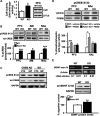
Figure 1.
Figure 1.
CREB activation and BDNF expression are increased in Rcan1 KO mice. A, CaN activity is elevated in the PFC of Rcan1 KO mice (p = 0.0259) and is not due to different protein levels of CaN (60 kDa). β-Tubulin (β-Tub), loading control. N = 9 KO, 6 WT. B, Enhanced pCREB S133 is seen in the PFC, AM, and NAc of Rcan1 KO mice. Total CREB levels are unchanged between genotypes. C, Identity confirmation of the pCREB signal used for quantification in this study. Viral-mediated CREB knockdown (KD) tissue from the cortex (ctx) and hippocampus (hip) were probed for pCREB S133 and reprobed for total (tot) CREB on the same blot. GAPDH, loading control. D, Acute blockade of CaN activity with FK506 eliminates the CREB activation differences between Rcan1 KO and WT mice. Pairwise comparisons of PFC percentage pCREB of WT-vehicle levels revealed a significant difference between WT and KO vehicle groups (p < 0.001) and no difference between KO-FK506 and WT-vehicle groups (p = 0.446) or between WT-FK506 and KO-FK506 groups (p = 1.000). N = 4 mice/group. The same effect was observed in the NAc. E, Bdnf mRNA (exon IV) and pro-BDNF protein levels (32 kDa) are increased in the PFC of Rcan1 KO mice. Semiquantitative PCR of cDNA synthesized from Bdnf mRNA bearing exon IV (confirmed with intron-spanning primers). N = 4 mice/genotype. Western blot of pro-BDNF levels. N = 4–6 mice/genotype. β-Actin mRNA levels and GAPDH staining confirms equal loading in each lane. *p < 0.05, **p < 0.01, ***p < 0.001.
Figure 2.
Figure 2.
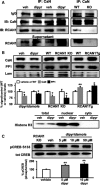
Disruption of RCAN1–CaN interaction alters subcellular phosphatase localization and leads to CREB activation. A, Treatment with 5 μm dipyridamole (dipyr) disrupts RCAN1 binding to CaN in the hippocampus. Immunoprecipitation of CaN shows reduced RCAN1 interaction with CaN following dipyridamole compared with vehicle treatment (veh). Signal specificity was confirmed using Rcan1 KO tissue. B, Dipyridamole treatment results in lower nuclear levels of CaN and its substrate, PP1. Genetic removal of RCAN1 (KO) also lowers nuclear levels of CaN and PP1. Conversely, overexpression of RCAN1 (Tg) increases nuclear CaN and PP1 levels. Equal loading and compartment fidelity confirmed with lamin A/C (Lam). Efficacy of subcellular localization protocol confirmed with histone H3 staining in total protein, nuclear, and cytoplasmic (cyto) fractions. Blots are representative of 2–3 replicates from three independent samples per treatment or genotype. C, Disruption of RCAN1-CaN with acute dipyridamole treatment activates CREB in the hippocampus, as measured by pCREB S133. Rcan1 KO tissue included for comparison. N = 5 vehicle, 5 WT+dipyr (5 μm), 4 WT+dipyr (10 μm). *p < 0.05, **p < 0.01.
Figure 3.
Figure 3.
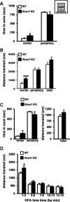
Rcan1 KO mice show decreased measures of anxiety in the OFA assay. A, Rcan1 KO mice spend more time in the center zone than the periphery of the OFA. B, Rcan1 KO mice travel a greater distance in the center zone without significant differences in total movement compared with WT littermates. C, Rcan1 KO mice also display greater center time when tested in a larger OFA (40 × 40 cm2). D, The increase in distance traveled by Rcan1 KO mice in the center zone occurs early during the test. Different cohorts used for data presented in A–D. A, B, N = 14 KO, 21 WT, time data for two WT samples lost during data collection. C, N = 8 KO, 9 WT. D, N = 11 KO, 11 WT. *p < 0.05, ***p < 0.001.
Figure 4.
Figure 4.
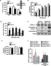
Rcan1 KO mice show decreased measures of anxiety in the EPM. A, Rcan1 KO mice spend significantly more time exploring the open arms of the EPM compared with their WT littermates. N = 10 KO, 12 WT. B, Rcan1 KO mice enter the open arms early in the EPM test (minute 1) whereas their WT littermates increased open-arm exploration starting at the third minute of testing compared with minute 1. N = 10 KO, 9 WT. C, Total distance moved and speed of Rcan1 KO mice are indistinguishable from WT mice in the EPM. N = 10 KO, 12 WT. D, Rcan1 KO mice display similar PPI of acoustic startle responses compared with their WT littermates. E, Western blot analysis of RCAN1 expression in the PFC of RCAN1 transgenic (Tg) mice used for this study. Upper blot is stained with an RCAN1 antibody that recognizes endogenously expressed RCAN1.1L (∼38 kDa) and RCAN1.4 (∼28 kDa) protein isoforms and transgenically expressed FLAG-tagged human RCAN1.1S protein (∼30 kDa; for more details, see Oh et al., 2005; Hoeffer et al., 2007). Lower blot is stained with a FLAG antibody, confirming expression of transgene. Nse-Cre driver line, CamkIIα-Cre (T-29) driver line. GAPDH, loading control. Image is representative of three independent blots. WT represents the WT control for each breeding cross. F, Mice overexpressing human RCAN1 early in development (Nse-RCAN1Tg1a) display decreased EPM open-arm time compared with WT littermates. No difference in open-arm time was observed driving an alternative RCAN1 construct (RCAN1Tg1) with the same Nse-Cre driver. No difference in open-arm time was also observed with postdevelopmental RCAN1 overexpression (CamkIIα-Cre) of either RCAN1 construct. WT-Tg1a (Nse) denotes control RCAN1Tg1a-only (no Cre) “WT” littermates from RCAN1Tg1a × Nse-Cre lines. WT-Tg1 (Nse) denotes control RCAN1Tg1-only (no Cre) “WT” littermates from RCAN1Tg1 × Nse-Cre lines. WT-Tg1 (CamkIIα) denotes control RCAN1Tg1a-only (no Cre) “WT” littermates from RCAN1Tg1a × CamkIIα-Cre lines. WT-Tg1 (CamkIIα) denotes control RCAN1Tg1-only (no Cre) “WT” littermates from RCAN1Tg1 × CamkIIα-Cre lines. N = 36 Nse-RCAN1Tg1a, 40 WT-Tg1a (Nse), 7 Nse-RCAN1Tg1, 11 WT-Tg1 (Nse), 32 CamkIIα-RCAN1Tg1a, 38 WT-Tg1a (CamkIIα), 11 CamkIIα-RCAN1Tg1, 17 WT-Tg1 (CamkIIα). For some datasets not normally distributed, Mann–Whitney analysis was used. *p < 0.05, ** or +p < 0.01, ***p < 0.001.
Figure 5.
Figure 5.
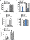
Acute pharmacological blockade of CaN rescues reduced anxiety in Rcan1 KO mice. A, Time in each OFA zone following intraperitoneal FK506 treatment. Vehicle-treated Rcan1 KO mice spend more time in the center zone than periphery of the OFA compared with similarly treated WT controls, whereas FK506-treated Rcan1 KO mice are not different from vehicle-treated WT controls. B, FK506 treatment reduces distance traveled by both WT and Rcan1 KO mice in all zones of the OFA. C, Movement in the OFA plotted as a ratio of distance traveled in each zone (zone distance) to total distance traveled during the test period. Using this measure, vehicle-treated Rcan1 KO mice move significantly more than vehicle-treated WT littermates in the center zone, whereas FK506-treated KO mice are indistinguishable from vehicle-treated WT mice. D, EPM open-arm and closed-arm time following CsA treatment via intraventricular cannulation. Pairwise comparisons (Dunn's with Bonferroni) revealed significant effects between the WT and KO vehicle groups (p = 0.014) and between the KO CsA and vehicle treatment groups (p = 0.004), while there was no difference between KO-CsA and WT-vehicle groups (p = 0.505) or WT-CsA groups (p = 0.995). Center zone measurements are not included but there is no difference between the groups. E, Total distance moved in the EPM is similar for WT and Rcan1 KO mice following intracerebroventricular administration of CsA or vehicle. OFA: N = 12 KO-vehicle, 20 WT-vehicle, 9 KO-FK506, 9 WT-FK506; EPM: N = 7 KO-vehicle, 11 WT-vehicle, 7 KO-CsA, 10 WT-CsA. **p < 0.01; ***p < 0.001; n.s., p > 0.05.
Figure 6.
Figure 6.
Rcan1 KO mice are resistant to the acute anxiogenic effects of SSRI administration. A, WT but not Rcan1 KO mice injected with intraperitoneal fluoxetine and tested 24 h later in the EPM show decreased open-arm time compared with their vehicle-treated (WT or KO) cohorts, indicating increased anxiety in fluoxetine-treated WT mice. B, Fluoxetine treatment does not change overall locomotor activity within or across genotypes. Total distance traveled for test period is shown. C, Open-arm time of EPM-naive mice following either 3 or 15 d of treatment with fluoxetine or vehicle. All animals tested had no prior experience with the EPM. Fluoxetine-treated Rcan1 KO mice increase time spent in the open arms, indicating reduced anxiety, compared with vehicle-treated KO mice after 3 d of treatment. After 15 d of treatment, fluoxetine-treated WT mice show a significant increase in open-arm time compared with WT-vehicle controls on day 3 or 15. Fluoxetine treatment also increased open-arm time in Rcan1 KO mice on day 15 compared with vehicle treatment, but the difference did not reach statistical significance. D, Total distance moved in the EPM by all the treatment groups is similar. No difference in movement was observed in EPM-naive animals tested after 1, 3, or 15 d of treatment. N (day 1, day 3, day 15) = (11, 9, 9) KO-vehicle; (12, 7, 8) WT-vehicle; (10, 9, 9) KO-fluoxetine; (11, 6, 6) WT-fluoxetine. WT-fluoxetine day 3 vs WT-day 15 fluoxetine denoted by *p < 0.05; **p < 0.01; *** or ‡p < 0.001; n.s., p > 0.05.

Similar articles

Cited by

References

    1. Alberts AS, Montminy M, Shenolikar S, Feramisco JR. Expression of a peptide inhibitor of protein phosphatase 1 increases phosphorylation and activity of CREB in NIH 3T3 fibroblasts. Mol Cell Biol. 1994;14:4398–4407. - PMC - PubMed
    1. Bahi A, Mineur YS, Picciotto MR. Blockade of protein phosphatase 2B activity in the amygdala increases anxiety- and depression-like behaviors in mice. Biol Psychiatry. 2009;66:1139–1146. doi: 10.1016/j.biopsych.2009.07.004. - DOI - PMC - PubMed
    1. Baldwin DS, Tiwari N. The pharmacologic treatment of patients with generalized anxiety disorder: where are we now and where are we going? CNS Spectr. 2009;14:5–12. - PubMed
    1. Barrot M, Olivier JD, Perrotti LI, DiLeone RJ, Berton O, Eisch AJ, Impey S, Storm DR, Neve RL, Yin JC, Zachariou V, Nestler EJ. CREB activity in the nucleus accumbens shell controls gating of behavioral responses to emotional stimuli. Proc Natl Acad Sci U S A. 2002;99:11435–11440. doi: 10.1073/pnas.172091899. - DOI - PMC - PubMed
    1. Baumgärtel K, Mansuy IM. Neural functions of calcineurin in synaptic plasticity and memory. Learn Mem. 2012;19:375–384. doi: 10.1101/lm.027201.112. - DOI - PubMed

Publication types

MeSH terms