. 2013 Oct 23;33(43):17089-94.
doi: 10.1523/JNEUROSCI.0522-13.2013.

piezo2b regulates vertebrate light touch response

Affiliations

piezo2b regulates vertebrate light touch response

Adèle Faucherre et al. J Neurosci. .

Abstract

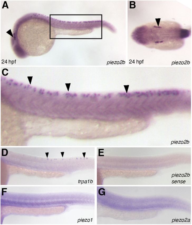
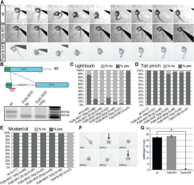
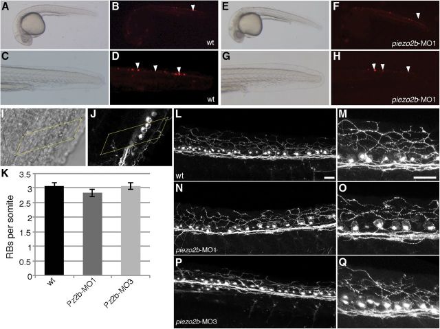
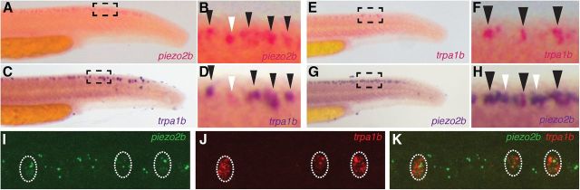
The sense of touch allows an organism to detect and respond to physical environmental stimuli. Mechanosensitive proteins play a crucial role in this process by converting the mechanical cue into a biological response. Recently, the Piezo family of stretch-activated ion channels has been identified as genuine mechanosensitive proteins. We set out to determine whether any of these genes are involved in touch response during zebrafish development. In situ hybridization indicates that piezo2b is specifically expressed in a subset of neurons (Rohon-Beard cells) responsible for detecting light touch. Using morpholino-mediated knockdown, we specifically targeted piezo2b and determined that it is involved in mediating touch-evoked response.

Keywords: Piezo2.


Figures

Figure 1.
Figure 1.
The expression pattern of piezo2b during zebrafish development. A–C, In situ hybridization analysis using an antisense piezo2b probe at 24 hpf. A, Piezo2b expression can be observed in the trigeminal ganglion (arrowhead) and Rohon–Beard cells (box). B, Dorsal view showing piezo2b expression in the trigeminal ganglion (arrowhead). C, Higher magnification of the boxed region in A. Piezo2b expression can be detected in the Rohon–Beard cells (arrowheads). E, To ensure that these observations were specific, we performed an identical assay using a sense piezo2b probe, which did not label anything. D, In situ hybridization analysis using an antisense trpa1b probe indicates that this gene is also expressed in Rohon–Beard neurons (arrowheads). F, G, The other two zebrafish piezo homologs, piezo1 and piezo2a, do not appear to be expressed in Rohon–Beard cells.
Figure 2.
Figure 2.
Piezo2b morphants are unresponsive to light touch but respond normally to mechanical and chemical stimuli. Touch response stimulation was performed on 24–27 hpf wild-type and piezo2b morphant embryos. A, wt panel, Time-lapse images of a wild-type embryo's response to touch-evoked stimuli. Piezo2b morphant embryos fail to respond to touch response stimulation (Pz2b MO panel). Trpa1b morphants respond normally to light touch (Trpa1b MO panel). B, RT-PCR analysis of Pz2b-MO3 morphant embryos. The Pz2b-MO3 targets the exon1/intron1 splice site. Primers designed to the start of exon1 and the end of exon 2 (arrows) are able to amplify the correct 270 bp fragment (top band) from wild-type embryo cDNA (lane 1). With morphant embryo cDNA, this band disappears, indicating that the correct splicing of piezo2b has been disrupted (excess primers form the lower band in all lanes). C, Quantitative analysis of wild-type, piezo2b, and trpa1b morphants subjected to touch response stimulation. Piezo2b morphants generated with morpholinos targeting the start codon (Pz2b-MO1), a nonoverlapping morpholino directed toward the 5′ UTR (Pz2b-MO2), or a splice-blocking morpholino targeting the exon1/intron1 boundary (Pz2b-MO3) fail to respond to tactile stimulation. The effect is enhanced slightly by increasing the concentration of morpholinos. Trpa1b morphants (Trpa1b-MO) respond normally in this assay (n = cumulative number of embryos from three independent assays). D, A tail pinch assay was performed on the same wild-type, piezo2b, and trpa1b morphants used in the previous light touch assay. The response of piezo2b and trpa1b morphants was indistinguishable from that of wild-type control embryos (n = cumulative number of embryos from three independent assays). E, Piezo2b morphants respond the same as control embryos when challenged with mustard oil. The assay was performed on the same wild-type and piezo2b morphants used in the previous light touch assay. However, mustard oil fails to elicit a response from trpa1b morphants (Trpa1b-MO; n = cumulative number of embryos from three independent assays). To analyze this phenotype in more detail, time-lapse images were recorded allowing us to count the number of writhings in response to mustard oil stimulation, an example of a wild-type embryo response is shown in F (black arrow indicates when a single writhe has been initiated). G, Piezo2b morphants respond to mustard oil with a similar number of writhings to that observed in wild-type embryos, while trpa1b morphants barely elicit a response to mustard oil (n = 10 embryos analyzed for each condition). Error bars indicate ±SEM. *p < 0.001, two-tailed Student's t test.
Figure 3.
Figure 3.
Rohon–Beard cells develop normally in piezo2b morphants. A–D, Rohon–Beard cells are clearly visible in wild-type embryos mosaically expressing mem-TdTomato under the control of the HuC promoter. A, Bright-field image of a wild-type embryo at 24 hpf mosaically expressing HuC:mem-TdTomato. B, The same embryo viewed with RFP-filtered UV light. The Rohon–Beard cells are indicated with arrowheads. C, D, The same embryo at higher magnification. The Rohon–Beard cells are indicated with arrowheads. E, Bright-field image of a piezo2b morphant embryo at 24 hpf mosaically expressing HuC:mem-TdTomato. F, The same embryo viewed with RFP-filtered UV light. The Rohon–Beard cells are indicated with arrowheads. G, H, The same embryo at higher magnification. The Rohon–Beard cells are indicated with arrowheads. Note: because the HuC promoter-driven mem-TdTomato expression is mosaic, this is not a quantitative assay and only illustrates that Rohon–Beard cells are present in piezo2b morphants. I–K, The average number of Rohon–Beard cells does not change following piezo2b knockdown. I, Bright-field image of a 24 hpf embryo immunolabeled with the zn-12 antibody; the yellow box delineates the somite boundaries. J, Fluorescent image of the same embryo. Three Rohon–Beard cell soma are clearly visible within the somite boundary (yellow box). K, Graph indicating the average number of Rohon–Beard cells (RB)/somite in wild-type (wt) embryos, and in two different piezo2b morphant groups (n = 6 embryos/condition, 5 somites/embryo). Error bar indicates ±SEM. Somite data were pooled, and no statistical difference was observed between these groups. L–Q, The gross morphology of Rohon–Beard soma and neurites is unaffected following piezo2b knockdown. L, Fluorescent image of a wild-type embryo labeled with the zn-12 antibody; peripheral neurites can be seen extending from individual Rohon–Beard soma to innervate the skin. Scale bar, 25 μm. M, The same image at a higher magnification. Scale bar, 25 μm. N–Q, Similar images taken from two different piezo2b morphant groups; the gross morphology of the Rohon–Beard soma and their neurites appears normal when compared with that of the wild-type controls (n = 6 embryos analyzed/condition).
Figure 4.
Figure 4.
A subset of piezo2b-expressing Rohon–Beard cells also express trpa1b. A–D, Two-color in situ hybridization with piezo2b labeled in red (A, B) and trpa1b in blue (C, D). Individual embryos were initially imaged to detect the red stain. Subsequently, following the blue stain, the same individual embryos were imaged again, allowing us to determine coexpression. All of the trpa1b-positive cells are also positive for piezo2b (B, D; black arrowheads); however, it is also possible to detect piezo2b-positive/trpa1b-negative Rohon–Beard cells (D; white arrowhead). E–H, A similar in situ hybridization assay performed with trpa1b in red (E, F) and piezo2b in blue (G, H). All of the trpa1b-positive cells are also positive for piezo2b (F, H; black arrowheads); however, it is also possible to detect piezo2b positive/trpa1b-negative Rohon–Beard cells (H; white arrowheads). I, J, Double-fluorescent in situ hybridization for piezo2b (I; green) and trpa1b (J; red). K, All trpa1b-positive cells also express piezo2b.

References

    1. Bhattacharya MR, Bautista DM, Wu K, Haeberle H, Lumpkin EA, Julius D. Radial stretch reveals distinct populations of mechanosensitive mammalian somatosensory neurons. Proc Natl Acad Sci U S A. 2008;105:20015–20020. doi: 10.1073/pnas.0810801105. - DOI - PMC - PubMed
    1. Brend T, Holley SA. Zebrafish whole mount high-resolution double fluorescent in situ hybridization. J Vis Exp. 2009;pii:1229. doi: 10.3791/1229. - DOI - PMC - PubMed
    1. Coste B, Mathur J, Schmidt M, Earley TJ, Ranade S, Petrus MJ, Dubin AE, Patapoutian A. Piezo1 and Piezo2 are essential components of distinct mechanically activated cation channels. Science. 2010;330:55–60. doi: 10.1126/science.1193270. - DOI - PMC - PubMed
    1. Eijkelkamp N, Linley JE, Torres JM, Bee L, Dickenson AH, Gringhuis M, Minett MS, Hong GS, Lee E, Oh U, Ishikawa Y, Zwartkuis FJ, Cox JJ, Wood JN. A role for Piezo2 in EPAC1-dependent mechanical allodynia. Nat Commun. 2013;4:1682. doi: 10.1038/ncomms2673. - DOI - PMC - PubMed
    1. Faucherre A, Pujol-Martí J, Kawakami K, López-Schier H. Afferent neurons of the zebrafish lateral line are strict selectors of hair-cell orientation. PLoS One. 2009;4:e4477. doi: 10.1371/journal.pone.0004477. - DOI - PMC - PubMed

Publication types

LinkOut - more resources