

GABA type B receptor signaling in proopiomelanocortin neurons protects against obesity, insulin resistance, and hypothalamic inflammation in male mice on a high-fat diet
- PMID: 24155320
- PMCID: PMC6618432
- DOI: 10.1523/JNEUROSCI.0897-13.2013
GABA type B receptor signaling in proopiomelanocortin neurons protects against obesity, insulin resistance, and hypothalamic inflammation in male mice on a high-fat diet
Abstract
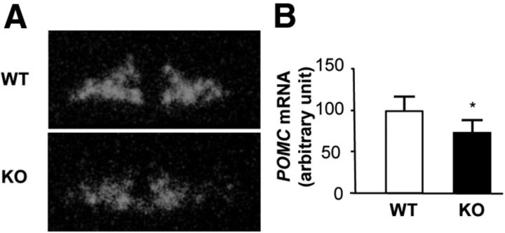
There is evidence suggesting that the GABA system in the arcuate nucleus, where orexigenic neuropeptide Y and agouti-related peptide as well as anorexigenic proopiomelanocortin (POMC) are expressed, plays an important role in energy balance. In this study, we generated POMC-specific GABAB receptor-deficient [knock-out (KO)] mice. Male KO mice on a high-fat diet (HFD) showed mild increases in body weight (BW) at the age of 9 weeks compared to wild-type (WT) mice, and the differences remained significant until 16 weeks old. However, there was no difference in BW in females between genotypes. While food intake was similar between genotypes, oxygen consumption was significantly decreased in the male KO mice. The insulin tolerance test revealed that the male KO mice were less insulin sensitive compared to WT mice at the age of 8 weeks, when there was no significant difference in BW between genotypes. Despite increased BW, POMC mRNA expression in the arcuate nucleus was significantly decreased in the KO mice compared to WT mice at the age of 16 weeks. Furthermore, the expression of TNFα as well as IL-6, proinflammatory markers in the hypothalamus, was significantly increased in the KO mice on a HFD compared to WT mice. This demonstrates that the deletion of GABAB receptors in POMC neurons in the male mice on a HFD results in obesity, insulin resistance, and hypothalamic inflammation. Furthermore, the decreased POMC expression in the obese KO mice suggests that the regulation of POMC expression through GABAB receptors is essential for proper energy balance.
Figures


Similar articles
-
AgRP Neuron-Specific Deletion of Glucocorticoid Receptor Leads to Increased Energy Expenditure and Decreased Body Weight in Female Mice on a High-Fat Diet.Endocrinology. 2016 Apr;157(4):1457-66. doi: 10.1210/en.2015-1430. Epub 2016 Feb 18. Endocrinology. 2016. PMID: 26889940
-
Overexpression of Smad7 in hypothalamic POMC neurons disrupts glucose balance by attenuating central insulin signaling.Mol Metab. 2020 Dec;42:101084. doi: 10.1016/j.molmet.2020.101084. Epub 2020 Sep 22. Mol Metab. 2020. PMID: 32971298 Free PMC article.
-
Hypothalamic Pomc expression restricted to GABAergic neurons suppresses Npy overexpression and restores food intake in obese mice.Mol Metab. 2020 Jul;37:100985. doi: 10.1016/j.molmet.2020.100985. Epub 2020 Apr 18. Mol Metab. 2020. PMID: 32311511 Free PMC article.
-
Food intake in early life and epigenetic modifications of pro-opiomelanocortin expression in arcuate nucleus.Mol Biol Rep. 2021 Apr;48(4):3773-3784. doi: 10.1007/s11033-021-06340-x. Epub 2021 Apr 20. Mol Biol Rep. 2021. PMID: 33877530 Review.
-
Mechanistic insight into high-fat diet-induced metabolic inflammation in the arcuate nucleus of the hypothalamus.Biomed Pharmacother. 2021 Oct;142:112012. doi: 10.1016/j.biopha.2021.112012. Epub 2021 Aug 10. Biomed Pharmacother. 2021. PMID: 34388531 Review.
Cited by
-
Deficiency of PTP1B Attenuates Hypothalamic Inflammation via Activation of the JAK2-STAT3 Pathway in Microglia.EBioMedicine. 2017 Feb;16:172-183. doi: 10.1016/j.ebiom.2017.01.007. Epub 2017 Jan 9. EBioMedicine. 2017. PMID: 28094236 Free PMC article.
-
Hypothalamic Inflammation: Is There Evidence for Human Obesity?Curr Obes Rep. 2014 Jun;3(2):242-7. doi: 10.1007/s13679-014-0104-0. Curr Obes Rep. 2014. PMID: 26626605
-
Epigenomic and metabolic responses of hypothalamic POMC neurons to gestational nicotine exposure in adult offspring.Genome Med. 2016 Sep 8;8(1):93. doi: 10.1186/s13073-016-0348-2. Genome Med. 2016. PMID: 27609221 Free PMC article.
-
Hypothalamic inflammation and gliosis in obesity.Curr Opin Endocrinol Diabetes Obes. 2015 Oct;22(5):325-30. doi: 10.1097/MED.0000000000000182. Curr Opin Endocrinol Diabetes Obes. 2015. PMID: 26192704 Free PMC article. Review.
-
Dietary sodium chloride attenuates increased β-cell mass to cause glucose intolerance in mice under a high-fat diet.PLoS One. 2021 Mar 17;16(3):e0248065. doi: 10.1371/journal.pone.0248065. eCollection 2021. PLoS One. 2021. PMID: 33730054 Free PMC article.
References
Publication types
MeSH terms
Substances
LinkOut - more resources
Full Text Sources
Other Literature Sources
Medical
Molecular Biology Databases
Research Materials
Miscellaneous