. 2014 Jan;88(1):303-13.
doi: 10.1128/JVI.02041-13. Epub 2013 Oct 23.

Minimal features of efficient incorporation of the hemagglutinin-neuraminidase protein into sendai virus particles

Affiliations

Minimal features of efficient incorporation of the hemagglutinin-neuraminidase protein into sendai virus particles

Manel Essaidi-Laziosi et al. J Virol. 2014 Jan.

Abstract

Two transmembrane glycoproteins form spikes on the surface of Sendai virus, a member of the Respirovirus genus of the Paramyxovirinae subfamily of the Paramyxoviridae family: the hemagglutinin-neuraminidase (HN) and the fusion (F) proteins. HN, in contrast to F, is dispensable for viral particle production, as normal amounts of particles can be produced with highly reduced levels of HN. This HN reduction can result from mutation of an SYWST motif in its cytoplasmic tail to AFYKD. HNAFYKD accumulates at the infected cell surface but does not get incorporated into particles. In this work, we derived experimental tools to rescue HNAFYKD incorporation. We found that coexpression of a truncated HN harboring the wild-type cytoplasmic tail, the transmembrane domain, and at most 80 amino acids of the ectodomain was sufficient to complement defective HNAFYKD incorporation into particles. This relied on formation of disulfide-bound heterodimers carried out by the two cysteines present in the HN 80-amino-acid (aa) ectodomain. Finally, the replacement of the measles virus H cytoplasmic and transmembrane domains with the corresponding HN domains promoted measles virus H incorporation in Sendai virus particles.


Figures
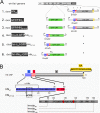
FIG 1
FIG 1
Schematic representation of rSeV genomes and of some HN protein features relevant to this study. (A) Outline of the parental SeV genome with its 6 genes flanked by the genomic promoter (GP) and the complement of the antigenomic promoter (AGPc). 1, Blow-up of the HN gene as it stands in rSeV-HNwt virus. The HN open reading frame (ORF) is flanked with gene start and end signals (gray bent arrows and lines) and with 3′ and 5′ untranslated sequences (UTR) (light gray thin tubes). In its ORF (wide colored tubes), the domains are indicated: cytoplasmic (c, in blue) carrying the SYWST motif, transmembrane (t, in red), ectodomain (e, in gray), and HA tag (HA, in yellow). 2, Outline of rSeV-GFP/HNwt virus, as in construct 1, with the rSeV genome containing an additional transcriptional unit between the M and F genes, encoding the green fluorescent protein (GFP) (in green). 3, Outline of rSeV-GFP/HNAFYKD virus, as in construct 2 except that the SYWST motif is replaced by AFYKD. 4, Outline of rSeV-hn-ct-GFP/HNAFYKD virus, as in construct 3 but with the GFP ORF fused at its N terminus to the hn cytoplasmic and transmembrane domains. 5, Outline of rSeV-hn-cmt-GFP/HNAFYKD virus, as in construct 4 but with the motif SYWST replaced by AFYKD in the hn cytoplasmic domain. 6, Outline of rSeV-hn-cte26-GFP/HNAFYKD virus. As in construct 4 but with the hn-ct domains extended with 26 amino acids of the hn ectodomain. Only highlighted parts (white on black) of the virus denominations were used for the annotation of the viruses in the figures and in Results. (B) Outline of the HN protein pointing to features important for this study (color code is as in panel A). HN is 593 amino acids long (576 aa + HA tag). Its wild-type (wt) cytoplasmic domain contains the 10SYWST14 motif, replaced by AFYKD as in HNAFYKD. α-HNc, outline of the cytoplasmic peptide sequence used to raise the α-HNc rabbit serum reacting against HN (wild type and mutated HNAFYKD). hn-cte26, hn-cte80, and hn-cte130, sequence additions with increasing extensions of the ectodomain fused to GFP, as in recombinant viruses analyzed in Fig. 2, 3, 4, and 6. The numbers in the denominations refer to the length of the ectodomain only, while the numbers in the figure refer to the total length form the HN N terminus. The cysteine residues (in red in the ectodmain, positions 129 and 138) refer to the cysteines changed to serines in experiments presented in Fig. 6 and 7.
FIG 2
FIG 2
First attempt to rescue incorporation of HNAFYKD in SeV particles. LLC-MK2 cells were infected with the indicated viruses described in Fig. 1A. (A) Western blot analysis of the cellular extracts (IC) and viral particles in the cell supernatants (VP) probed with α-HNSDS,-MSDS, -NSDS, and -FSDS (upper panel), α-HNc (middle panel), and α-GFP (lower panel) antibodies. GFP (black arrowhead) and hn-ct-, hn-cmt-, or hn-cte26-GFP proteins (*) are indicated. (B) Infected cells were radiolabeled with [35S]methionine and cysteine from 16 to 24 h postinfection. Surface (S) and total (T) immunoprecipitations (IP) were performed using α-GFP or α-HA as indicated. Lane V, 35S-SeV labeled viral proteins loaded as protein markers. Immunopreciptated GFP (green arrow) and hn-ct- or hn-cte-GFP proteins (*) are indicated. (C) Eighteen hours postinfection, LLC-MK2 cells infected with the indicated viruses were radio-pulse-labeled for 10 or 20 min to control synthesis. After the 20-min pulse, the cells were further chased for 90 min. The GFP-containing proteins were then immunoprecipitated using α-GFP and directly analyzed by PAGE and detected by autoradiography. (D) LLC-MK2 cells grown on coverslips were infected with the rSeVs expressing the hn-ct's and hn-cte26 fused to GFP as described for panel A. At 24 hours postinfection, the cells were fixed, stained with DAPI, and observed by confocal microscopy. GFP proteins, green; nuclei, blue.
FIG 3
FIG 3
Second attempt to rescue incorporation of HNAFYKD in SeV particles. (A) Schematic representation of rSeV viruses encoding hn-cte26-GFP, hn-cte80-GFP, and hn-cte130-GFP. Note that the hn-cte-GFPs are all in the wt (SYWST) configuration and that the endogenous HN is in its mutated (AFYKD) one. The same color code as for Fig. 1A is used. (B) LLC-MK2 cells were infected with the indicated viruses, and Western blot analysis of the cellular extracts (IC) and the viral particles in the cell supernatant, probed with the indicated antibody preparations, are presented. *, hn-cte26-GFP protein detected with α-HNSDS, α-HNc, and α-GFP antibodies at the expected molecular mass (all panels). α-HNc recognizes both full HN and GFP HN tails. ?, potential hn-cte80-GFP and hn-cte130-GFP proteins detected using α-HNc antibody but with an unexpected molecular mass (middle panel). (C) HNAFYKD incorporation in VP after infections with the indicated viruses, quantified (using ImageJ software) in relation to N and normalized to incorporation of wild-type HN (lane 2 of panel B) taken as 100%. Data were obtained from three separate experiments.
FIG 4
FIG 4
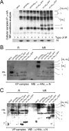
Study of endogenous HN oligomerization in the presence of hn-cte26-GFP, hn-cte80-GFP, and hn-cte130-GFP. LLC-MK2 cells were infected with the indicated viruses as for Fig. 3. Cellular extracts were collected at 24 h postinfection and subjected to surface (S) or total (T) immunoprecipitations using α-HA antibody. Viral particles were purified form the cell supernatants. (A) Western blot analysis of the cellular immunoprecipitates performed under nonreducing (NR) conditions and probed with α-HNc (upper panel). N was recovered from the same samples with α-N antibody as a control for surface IP (lower panel). (B) Western blot analysis of the purified viral particles performed under reducing (R) and nonreducing (NR) conditions probed with α-HNc antibody. (C) Exactly as in panel B except for 17.5% SDS-PAGE, as opposed to 10% in panel B, and for use of a higher-sensitivity ECL (Pierce ECL Plus).
FIG 5
FIG 5
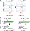
Sequence comparison of hn-cte-GFPs in plasmids and rescued viruses. (A) Viral RNA was recovered from recombinant virus particles, and the genes encoding hn-cte80-GFP and hn-cte130-GFP were reverse transcribed (RT) using a primer of positive polarity positioned in the 5′ extremity of the M gene. The RT products were then amplified by PCR using primers flanking the additional transcription units carrying the hn-cte-GFP genes situated between the M and F genes. RT-PCR products were sequenced and compared to the sequence of the original cDNA used for virus rescue. Highlighted nucleotides in gray indicate the G-to-T and the G-to-A substitutions leading to the insertion of stop codons in the hn-cte80-GFP and hn-cte130-GFP ORFs, respectively. (B) Schematic representation of hn-cte80-GFP and hn-cte130-GFP proteins theoretically expressed by the full-length cDNA plasmid used for virus rescue (plasmid) and the proteins actually expressed by the recovered viruses (virus).
FIG 6
FIG 6
Importance of disulfide bond formation for HNAFYKD rescue in viral particles. (A) Schematic representation of the two hn-cte80 proteins harboring SYWST or AFYKD (hn-cte80 and hn-cmte80), each presented with the normal cysteine composition at positions 69 and 78 of the ectodomain or with the replacement of the 2 cysteines (C/C) by 2 serines (S/S). The 4 rSeVs express HA-HNAFYKD, as endogenous HN. The color code is as in Fig. 1A. (B and C) LLC-MK2 cells were infected with the viruses described in panel A and Fig. 1A. (B) Western blot analysis of the cellular extracts (IC) and viral particles present in the cell supernatants (VP) probed with α-HNSDS,-MSDS, -NSDS, and -FSDS (upper panel) and α-HA (lower panel). C and S indicate the presence of cysteine or serine, respectively, at position 69 or 78 of the ectodomain of hn-cte80 and hncmte80. (C) Left panels, Western blot analysis of immunoprecipitations (IP) performed with α-HA antibody under nonreducing (NR) (upper panel) and reducing (R) (lower panel) conditions and probed with α-HNc. Right panels, similar Western blot analysis of the corresponding viral particles (VP) resuspended in a nonreducing sample buffer. (D). Western blots for the experiment presented in panel B (VP) plus two similar independent experiments were quantified using ImageJ software for estimation of HN incorporation in viral particles. In each lane HN was normalized to N, and the result of HNwt incorporation was taken as 100%.
FIG 7
FIG 7
Cysteines 69 and 78 are not required for HNwt incorporation in viral particles. (A) Schematic representation of rSeV-GFP/HN-C2S (construct 3), in which HN harbors replacements of cysteines 69 and 78 by serine. (B) Western blots of cellular extracts (IC, left panels) and supernatant (VP, right panels) from noninfected (M) LLC-MK2 or cells infected with the indicated viruses analyzed under reducing conditions (R) (upper panel) using α-HNSDS,-MSDS, -NSDS, and -FSDS and under nonreducing (NR) (lower panel) conditions using α-HNc. (C) Quantification of HN incorporation into VP. Western blots were quantified using ImageJ software, and HN was normalized to N content. Incorporation of HNwt was taken as 100%. Data were obtained from three independent experiments.
FIG 8
FIG 8
Incorporation of MeV H into SeV particles. (A) Schematic representation of the rSeVs expressing MeV H. As shown in construct 4, hnctH refers to MeV H in which the cytoplasmic and transmembrane regions have been replaced with the corresponding domains of SeV HN. (B) LLC-MK2 cells were infected with the indicated viruses. The infected cells were radiolabeled with [35S]methionine-cysteine at 16 h postinfection and were collected 24 h later. Upper panels, SDS-PAGE autoradiogaphy of cellular extracts (IC) and virus particles (VP). Middle panel, Western blot analysis of the SDS-PAGE probed with α-HA. Lower panel, SDS-PAGE autoradiography of immunoprecipitates performed with α-MeV H on total cellular extracts and VP prepared as for panel A. Note that MeV H and HN exhibit the same migration profile. (C) LLC-MK2 cells were mock infected (lanes M) or infected with rSeV-GFP/HNAFYKD (lanes 2), rSeV-MeV H/HNAFYKD (lanes 3), or rSeV-MeV hnctH/HNAFYKD (lanes 4) viruses as described for panel A. Sixteen hours postinfection, cells were [35S]methionine-cysteine radiolabeled for 24 h. Cellular extracts were directly analyzed under reducing conditions (IC/R) or immunoprecipitated using an α-MeV H antibody (IP) and analyzed under reducing (R) or nonreducing (NR) conditions. Lower panels, Western blot analysis of the cellular extracts (IC) and of the immunoprecipitates probed with α-HA.
FIG 9
FIG 9
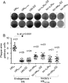
Infectivity tests of recombinant rSeV viruses. LLC-MK2 cells grown in six-well petri dishes were infected with the indicated viruses (for their description, see Fig. 3 and 6) at appropriate dilutions. After 1 h of adsorption, infected cells were incubated for 5 days at 33°C under conditions of infectious virus titration. Cells were then fixed with trichloroacetic acid (5% in H2O) for 10 min at room temperature and stained with crystal violet. (A) Images of stained wells showing the plaques. (B) Pictures of the well were taken to allow measurement of the plaque size using ImageJ software. In each well, the plaque size was normalized to the size of the well. n, number of plaques measured for each recombinant virus.

Similar articles

Cited by

References

    1. Lamb RA, Parks GD. 2007. Paramyxoviridae: the viruses and their replication, p 1449–1496 In Knipe DM, Howley PM. (ed), Fields virology, 5th ed, vol 1 Lippincott Williams & Wilkins, Philadelphia, PA
    1. Blumberg B, Giorgi C, Roux L, Raju R, Dowling P, Chollet A, Kolakofsky D. 1985. Sequence determination of the Sendai virus HN gene and its comparison to the influenza virus glycoproteins. Cell 41:269–278. 10.1016/0092-8674(85)90080-7 - DOI - PubMed
    1. Markwell MA, Fox CF. 1980. Protein-protein interactions within paramyxoviruses identified by native disulfide bonding or reversible chemical cross-linking. J. Virol. 33:152–166 - PMC - PubMed
    1. Vidal S, Mottet G, Kolakofsky D, Roux L. 1989. Addition of high-mannose sugars must precede disulfide bond formation for proper folding of Sendai virus glycoproteins. J. Virol. 63:892–900 - PMC - PubMed
    1. Bose S, Welch BD, Kors CA, Yuan P, Jardetzky TS, Lamb RA. 2011. Structure and mutagenesis of the parainfluenza virus 5 hemagglutinin-neuraminidase stalk domain reveals a four-helix bundle and the role of the stalk in fusion promotion. J. Virol. 85:12855–12866. 10.1128/JVI.06350-11 - DOI - PMC - PubMed

Publication types

LinkOut - more resources