. 2014 Jan;88(1):314-24.
doi: 10.1128/JVI.02077-13. Epub 2013 Oct 23.

CD4 T cell help is limiting and selective during the primary B cell response to influenza virus infection

Affiliations

CD4 T cell help is limiting and selective during the primary B cell response to influenza virus infection

Shabnam Alam et al. J Virol. 2014 Jan.

Abstract

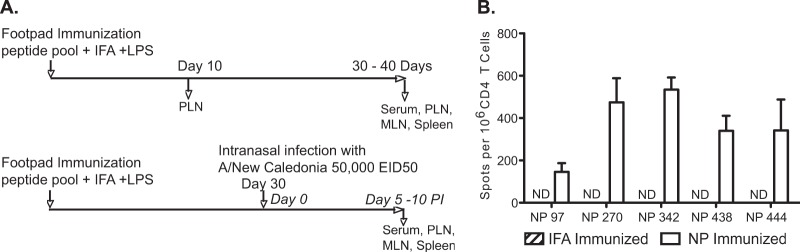
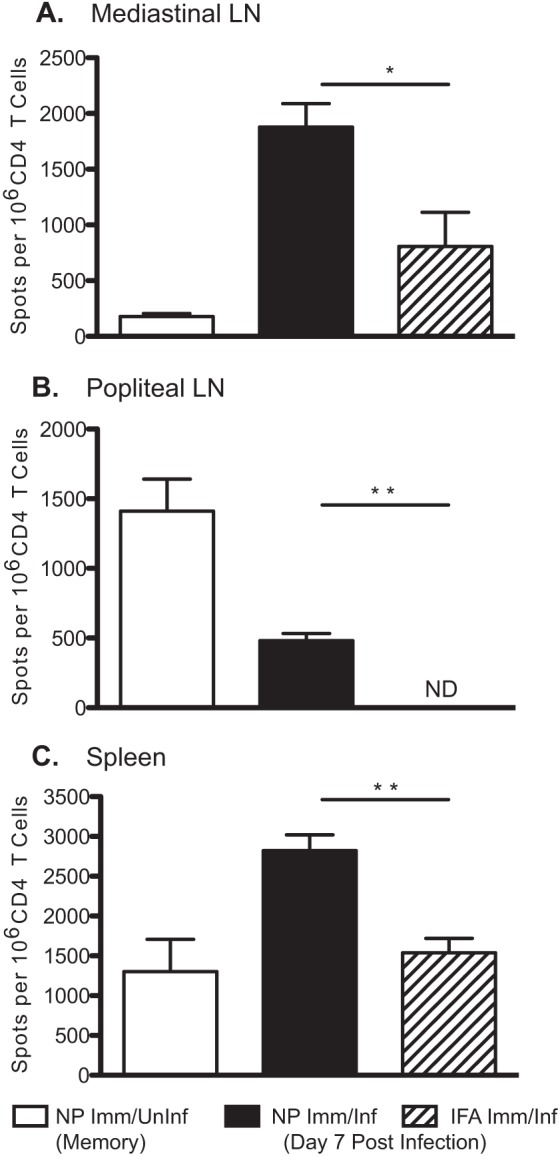
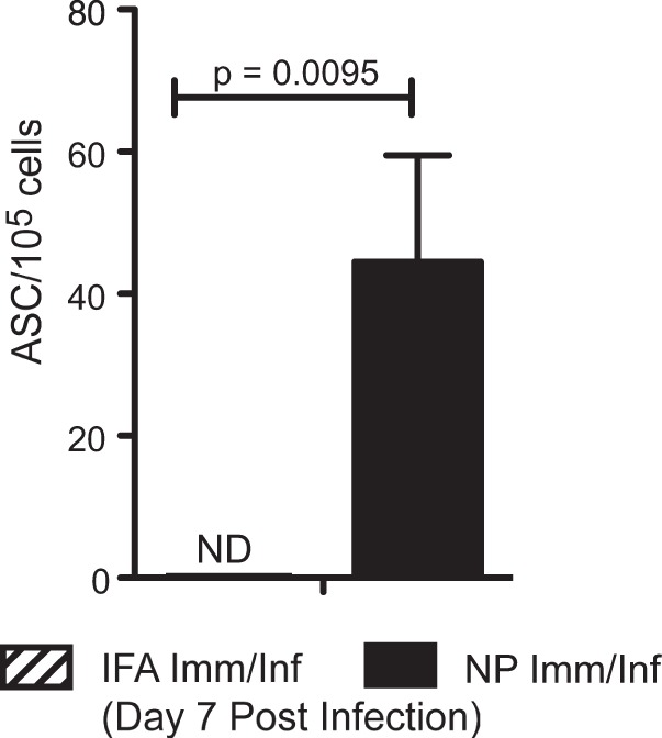
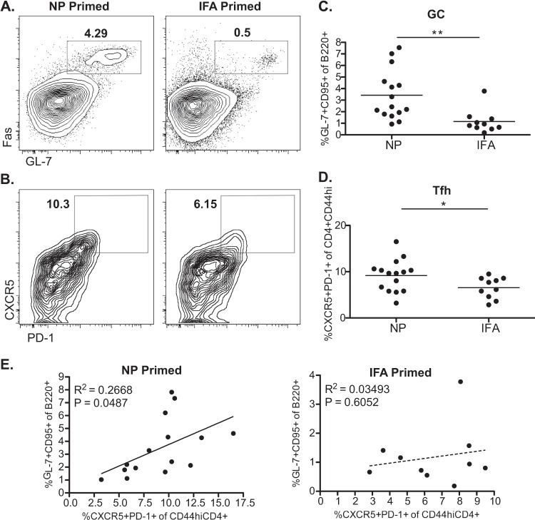
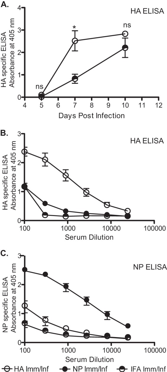
Influenza virus vaccination strategies are focused upon the elicitation of protective antibody responses through administration of viral protein through either inactivated virions or live attenuated virus. Often overlooked in this strategy is the CD4 T cell response: how it develops into memory, and how it may support future primary B cell responses to heterologous infection. Through the utilization of a peptide-priming regimen, this study describes a strategy for developing CD4 T cell memory with the capacity to robustly expand in the lung-draining lymph node after live influenza virus infection. Not only were frequencies of antigen-specific CD4 T cells enhanced, but these cells also supported an accelerated primary B cell response to influenza virus-derived protein, evidenced by high anti-nucleoprotein (NP) serum antibody titers early, while there is still active viral replication ongoing in the lung. NP-specific antibody-secreting cells and heightened frequencies of germinal center B cells and follicular T helper cells were also readily detectable in the draining lymph node. Surprisingly, a boosted memory CD4 T cell response was not sufficient to provide intermolecular help for antibody responses. Our study demonstrates that CD4 T cell help is selective and limiting to the primary antibody response to influenza virus infection and that preemptive priming of CD4 T cell help can promote effective and rapid conversion of naive B cells to mature antibody-secreting cells.


Figures

FIG 1
FIG 1
Immunodominant influenza virus epitopes prime a robust CD4 T cell response. (A) Experimental protocol used throughout this study. To study the effects of memory on the response to subsequent infection, SJL mice were immunized in the footpad with an emulsion of peptides in IFA and LPS, as described in Materials and Methods, and were either tested directly 10 days postimmunization or rested for 4 weeks for studies on infection. One cohort of these mice was not infected, and a second cohort was infected intranasally with 5 × 10⁴ EID50 A/New Caledonia/20/99 at 30 days postimmunization. (B) To determine the primary response to peptides used for priming, CD4 T cells were isolated from the draining popliteal lymph nodes (PLN) 10 days after immunization. CD4 T cells from NP- and control IFA-immunized mice were recalled with NP peptides used in immunization. The data are presented as mean spots per million CD4 T cells from three individual mice and are representative of three experiments. ND, no detectable response. Error bars indicate standard deviations.
FIG 2
FIG 2
Influenza virus infection rapidly expands peptide-primed CD4 T cell memory. NP peptide- or IFA-immunized mice were challenged intranasally at 4 weeks postimmunization with 5 × 10⁴ EID50 A/New Caledonia/20/99. On day 7 postinfection, CD4 T cells were isolated from MLN (A), PLN (B), and spleen (C) and tested in an IFN-γ ELISPOT assay for reactivity to the pool of NP peptides. Experimental groups included NP peptide-immunized and infected mice (Imm/Inf) and IFA-immunized and infected mice. Uninfected NP-peptide immunized mice (Imm/Uninf) were assayed as a control for memory at day 0 of infection (day 30 postimmunization). The data are presented as mean spots per million CD4 T cells from 3 to 5 mice per group and are representative of at least three experiments. ND, no detectable response for IFA-primed PLN. Error bars indicate standard deviations. *, P < 0.01; **, P < 0.005, Student's t test.
FIG 3
FIG 3
CD4 T cell help potentiates future serum antibody response to infection. Mice were primed and infected as illustrated in Fig. 1A. Sera were collected from the indicated cohorts of naive, NP peptide-immunized (infected or uninfected), and IFA-immunized and infected mice and tested for reactivity toward recombinant NP protein. NP protein-specific IgG was measured via ELISA. (A) Kinetics of NP-specific antibody accumulation in sera collected at three different time points: day 5, day 7, and day 10. Absorbance readings were made at a dilution 1:900, mean values from 2 to 5 individual mice are shown. (B) Serial dilutions (3-fold starting at 1:100) of sera from day 7 postinfection indicating fold increase in NP-specific IgG titers. The inverse dilution is shown. NP-specific IgG was ∼150-fold higher in NP peptide-primed and infected mice than in IFA-primed and infected mice. Data are shown as mean values from five individual mice, representative of three separate experiments. Error bars indicate standard deviations. *, P < 0.02; ***, P < 0.0001, Student's t test.
FIG 4
FIG 4
Accelerated NP protein-specific B cell response to infection in peptide-primed and infected animals. On day 7 postinfection, cells were isolated from the MLN of NP- peptide-immunized and infected mice and IFA-immunized and infected mice. Antibody-secreting cells were tested for reactivity toward recombinant NP protein from the A/New Caledonia/20/99 virus and detected by ELISPOT assay. IgG was measured with IgG-specific secondary antibody reagent. Data shown depict 5 mice per group, representative of three independent experiments. Error bars indicate standard deviations. ND, no detectable response for IFA-primed and infected mice.
FIG 5
FIG 5
Accelerated primary B cell response characterized by day 7 germinal center B cell and Tfh cell populations. Single-cell suspensions prepared from the MLN of an NP peptide pool or IFA-immunized mice were stained and analyzed for the frequency of GC B cells (B220+ GL-7+ Fas+) (A) and Tfh cells (CXCR5++ PD-1++ CD4+ CD44hi) (B). Representative FACS plots are shown. (C and D) Plots depicting frequencies of GC B cells and Tfh cells show the mean value for 10 (IFA) to 15 (NP) individual mice per group, combined from two separate experiments. GC B cell frequency is indicated as a percentage of the B220+ cell population. Tfh cell frequency is indicated as percentage of the CD4+ CD44hi cell population. *, P < 0.05; **, P < 0.01, Student's t test. (E) Correlation of Tfh cell frequency and GC B cell frequency in NP-primed or IFA-primed mice. Pearson test was performed assuming Gaussian distribution, and R2 and P values are shown.
FIG 6
FIG 6
Provision of CD4 T cell help is selective for antibody responses of intramolecular specificity. Serum antibody responses to HA were determined in a manner similar to that for the ELISAs described in the legend of Fig. 3. Mice were HA peptide primed and then challenged with infection, with serum levels of HA-specific IgG evaluated on days 5, 7, and 10 postinfection. A serum dilution of 1:900 was used, and mean values from 2 to 5 individual mice, representative of three separate experiments, are shown (A). Peptide priming with influenza virus-derived epitopes boosts the serum antibody response of intramolecular-specific B cells for both HA-specific (B) and NP-specific (C) responses. Inverse 3-fold serum dilutions starting at 1:00 from primed and day 7 infected animals are shown. Data are shown as mean values from five individual mice, representative of three separate experiments. Error bars indicate standard deviations. ns, not significant; *, P < 0.05, Student's t test.

References

    1. Nichol KL, Treanor JJ. 2006. Vaccines for seasonal and pandemic influenza. J. Infect. Dis. 194(Suppl 20):S111–S118. 10.1086/507544 - DOI - PubMed
    1. Osterholm MT, Kelley NS, Sommer A, Belongia EA. 2012. Efficacy and effectiveness of influenza vaccines: a systematic review and meta-analysis. Lancet Infect. Dis. 12:36–44. 10.1016/S1473-3099(11)70295-X - DOI - PubMed
    1. Jackson LA, Gaglani MJ, Keyserling HL, Balser J, Bouveret N, Fries L, Treanor JJ. 2010. Safety, efficacy, and immunogenicity of an inactivated influenza vaccine in healthy adults: a randomized, placebo-controlled trial over two influenza seasons. BMC Infect. Dis. 10:71. 10.1186/1471-2334-10-71 - DOI - PMC - PubMed
    1. Treanor JJ, Talbot HK, Ohmit SE, Coleman LA, Thompson MG, Cheng PY, Petrie JG, Lofthus G, Meece JK, Williams JV, Berman L, Breese Hall C, Monto AS, Griffin MR, Belongia E, Shay DK. 2012. Effectiveness of seasonal influenza vaccines in the United States during a season with circulation of all three vaccine strains. Clin. Infect. Dis. 55:951–959. 10.1093/cid/cis574 - DOI - PMC - PubMed
    1. Weinfurter JT, Brunner K, Capuano SV, III, Li C, Broman KW, Kawaoka Y, Friedrich TC. 2011. Cross-reactive T cells are involved in rapid clearance of 2009 pandemic H1N1 influenza virus in nonhuman primates. PLoS Pathog. 7:e1002381. 10.1371/journal.ppat.1002381 - DOI - PMC - PubMed

Publication types

MeSH terms