

Primary central nervous system T-cell lymphoma mimicking meningoencephalomyelitis in a cat
- PMID: 24155454
- PMCID: PMC3659459
Primary central nervous system T-cell lymphoma mimicking meningoencephalomyelitis in a cat
Abstract
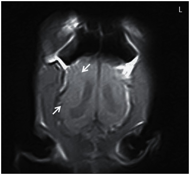
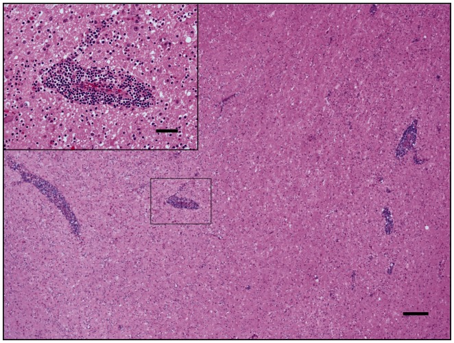
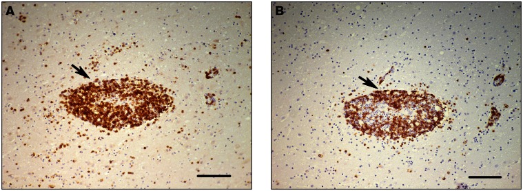
A cat was presented with right head tilt and circling. The lack of expression of virus antigens did not support the postmortem diagnosis of encephalomyelitis pointing to a diffuse primary central nervous system T-cell lymphoma on the basis of CD3 and CD45R co-expression with absence of CD79α staining.
Lymphome primaire de système nerveux central type T imitant méningo-encéphalomyélite chez un chat. Un chat est venu avec inclinaison de la tête à droite et circling. L’absence d’expression des antigènes du virus ne prend pas en charge le diagnositic post mortem e d’une encéphalomyélite pointant vers un lymphome primaire du système nerveux central type T diffus sur la base de CD3 et CD45R coexpression avec absence CD79α expression.(Traduit par les auteurs).
Figures
Similar articles
-
Periventricular spread of primary central nervous system T-cell lymphoma in a cat.J Comp Pathol. 2009 Jan;140(1):54-8. doi: 10.1016/j.jcpa.2008.09.003. Epub 2008 Dec 3. J Comp Pathol. 2009. PMID: 19056092
-
Central diabetes insipidus in a cat with central nervous system B cell lymphoma.J Feline Med Surg. 2011 Oct;13(10):787-92. doi: 10.1016/j.jfms.2011.07.005. Epub 2011 Sep 8. J Feline Med Surg. 2011. PMID: 21906986 Free PMC article.
-
Pathology in practice. Cutaneous, nonepitheliotropic T-cell lymphoma.J Am Vet Med Assoc. 2012 Oct 15;241(8):1035-7. doi: 10.2460/javma.241.8.1035. J Am Vet Med Assoc. 2012. PMID: 23039977 No abstract available.
-
Diagnostic immunohistochemistry of primary and secondary central nervous system neoplasms of dogs and cats.J Vet Diagn Invest. 2024 Mar;36(2):153-168. doi: 10.1177/10406387231221858. Epub 2024 Jan 17. J Vet Diagn Invest. 2024. PMID: 38234003 Free PMC article. Review.
-
Primary reticulosis of the central nervous system.Vet Clin North Am Small Anim Pract. 1980 Feb;10(1):57-63. doi: 10.1016/s0195-5616(80)50003-3. Vet Clin North Am Small Anim Pract. 1980. PMID: 6996289 Review. No abstract available.
Cited by
-
Neuropathology of Central and Peripheral Nervous System Lymphoma in Dogs and Cats: A Study of 92 Cases and Review of the Literature.Animals (Basel). 2023 Feb 27;13(5):862. doi: 10.3390/ani13050862. Animals (Basel). 2023. PMID: 36899719 Free PMC article.
-
CD56+ B-cell Neurolymphomatosis in a Cat.J Comp Pathol. 2019 May;169:25-29. doi: 10.1016/j.jcpa.2019.03.004. Epub 2019 May 13. J Comp Pathol. 2019. PMID: 31159947 Free PMC article.
-
Flow cytometric features of lymphoid subsets in healthy and diseased cats.Front Vet Sci. 2025 Aug 1;12:1640229. doi: 10.3389/fvets.2025.1640229. eCollection 2025. Front Vet Sci. 2025. PMID: 40822655 Free PMC article.
-
Apparent diffusion coefficient value for a B-cell central nervous system lymphoma in a cat.JFMS Open Rep. 2018 Jan 23;4(1):2055116917750762. doi: 10.1177/2055116917750762. eCollection 2018 Jan-Jun. JFMS Open Rep. 2018. PMID: 29383265 Free PMC article.
-
Primary nervous system lymphoma in cats.J Vet Diagn Invest. 2022 Jul;34(4):712-717. doi: 10.1177/10406387221090281. Epub 2022 Apr 20. J Vet Diagn Invest. 2022. PMID: 35442117 Free PMC article.
References
-
- Summers BA, Cummings JF, De Lahunta A. Tumors of the central nervous system. In: Summers BA, Cummings JF, De Lahunta A, editors. Veterinary Neuropathology. 1st ed. St. Louis, Missouri: Mosby; 1995. pp. 379–380.
-
- Koestner A, Higgins RJ. Tumors of the nervous system. In: Meuten DJ, editor. Tumors in Domestic Animals. 4th ed. Ames, Iowa: Iowa State University Press; 2002. pp. 697–738.
-
- Morrison LR, Freel K, Henderson I, Hahn C, Smith SH. Lymphoproliferative disease with features of lymphoma in the central nervous system of a horse. J Comp Pathol. 2008;139:256–261. - PubMed
-
- Palus V, Volk AH, Lamb CR, Targett MP, Cherubini GB. MRI Features of CNS lymphoma in dogs and cats. Vet Radiol Ultrasound. 2011;53:44–49. - PubMed
-
- Nakamoto Y, Ozawa T, Uchida K, Omori K, Hase K, Nakaichi M. Primary intra-axial B-cell lymphoma in a cat. J Vet Med Sci. 2008;71:207–210. - PubMed
Publication types
MeSH terms
LinkOut - more resources
Full Text Sources
Research Materials
Miscellaneous