

Smear Layer Evaluation on Root Canal Preparation with Manual and Rotary Techniques using EDTA as an Irrigant: A Scanning Electron Microscopy Study
- PMID: 24155580
- PMCID: PMC3768081
Smear Layer Evaluation on Root Canal Preparation with Manual and Rotary Techniques using EDTA as an Irrigant: A Scanning Electron Microscopy Study
Abstract
Introduction: The aim of any root canal treatment is to achieve a canal free of micro organisms, residual pulp remnants, debris and smear layer for the long term success of the procedure. Manual and automated instrumentation techniques along with proper irrigation regime is used to arrive at the aforementioned goal. Many authors focused on the preparation capabilities of various manual and rotary instruments but very few investigators stressed on the actual cleaning abilities of these instruments.
Aims and objectives: This study was undertaken to evaluate the cleaning efficiency of manual K flex files and rotary Pro File systems in the root canals using a scanning electron microscope.
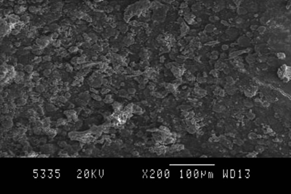
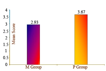
Material and methods: Thirty single rooted mandibular first premolars were divided into two groups and randomized (the manual group-M and the ProFile group-P) with respect to the preparation technique. The Manual group was hand instrumented with stainless steel K- Flexofiles by means of a conventional filing technique. The Pro File group was instrumented according to the manufacturer's instructions using a rotary handpiece. All canals were shaped and cleaned under frequent irrigation with EDTA. Final irrigation was carried out with 3 mL of normal saline solution to neutralize the action of the irrigant. The roots were split, one half of each tooth was selected for further SEM technique analysis and examined under the scanning electron microscope. The canal walls were quantitatively evaluated for the amount of debris and smear layer. The apical, middle and coronal regions of the canal surface, were graded (1-5) for debris and smear layer. A statistical analysis was performed using a Mann-Whitney Rank Sum test. ProFile performed least effective cleaning. Manual K-Flexofiles led to a grooved pattern.
Results and conclusion: A statistically significant difference was observed (p<0.05) between the two instrumentation techniques concerning the amount of debris and smear layer at the apical level. The manually filed canals had less debris and smear layer than those using a rotary technique. It was concluded from this study that none of the instrumentation techniques employed, produced the canal walls which were free of surface debris and smear layer. The manual instrumentation technique was better in cleaning the canals compared to the ProFile rotary Ni-Ti instruments despite the step-back technique used for manual instrumentation. How to cite this article: Manjunatha M, Kini A, Sudhakar V, Sunil K V C, Hiremath V K, Shah A. Smear Layer Evaluation on Root Canal Preparation with Manual and Rotary Techniques using EDTA as an Irrigant: A Scanning Electron Microscopy Study. J Int Oral Health 2013; 5(1):66-78.
Keywords: Clinical research; Oral Health; Tooth loss.
Conflict of interest statement
Conflict of Interest: None Declared
Figures


References
-
- Bolanos OR, Jensen JR. Scanning Electron Microscopic comparisons of the efficacy of various methods of root canal preparation. J Endod. 1980;6:815–822. - PubMed
-
- Haikel Y, Allemann C. Effectiveness of four methods for preparing root canals; a scanning electron microscopic evaluation. J Endod. 1988;14:340–345. - PubMed
-
- Hulsmann M, Rummelin C, Schafers F. Root canal cleanliness after preparation with different endodontic handpieces and hand instruments; a comparative scanning electron microscopy investigation. J Endod. 1977;23:301–306. - PubMed
-
- Versumer J, Hulsmann M, Schafers F. A comparative study of root canal preparation using ProFile .04 and Lightspeed rotary Ni-Ti instruments. Int Endod J. 2002;35:37–46. - PubMed
-
- Pashley DH, Michelich V, Kehl T. Dentin permeability; effects of smear layer removal. J Prosthet Dent. 1981;46:531–537. - PubMed
LinkOut - more resources
Full Text Sources