

Silencing of atp6v1c1 prevents breast cancer growth and bone metastasis
- PMID: 24155661
- PMCID: PMC3805834
- DOI: 10.7150/ijbs.6030
Silencing of atp6v1c1 prevents breast cancer growth and bone metastasis
Abstract
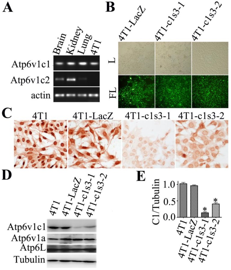
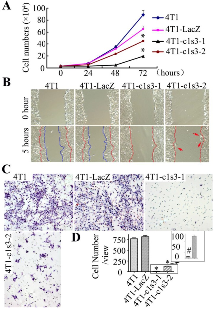
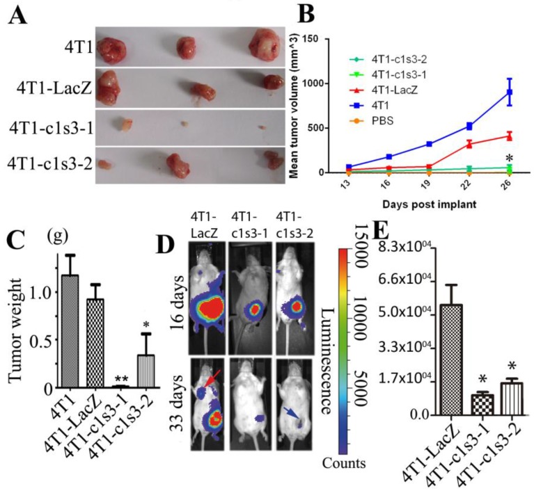
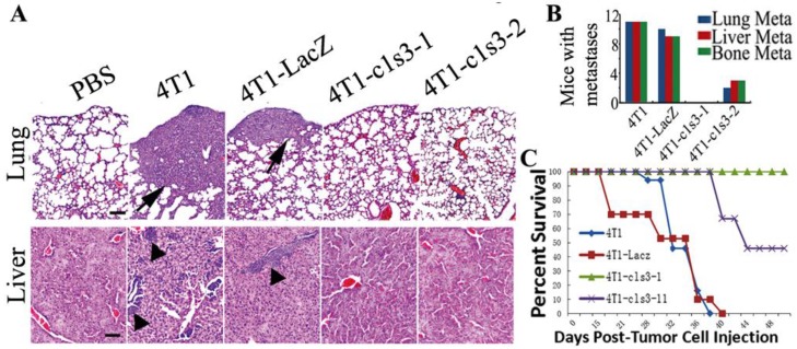
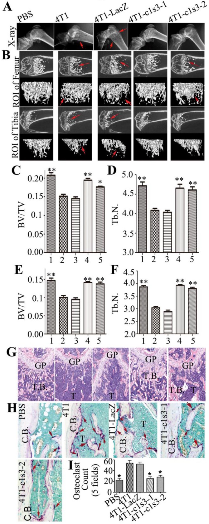
Previous studies have shown that Atp6v1c1, a regulator of the assembly of the V0 and V1 domains of the V-ATPase complex, is up-regulated in metastatic oral tumors. Despite these studies, the function of Atp6v1c1 in tumor growth and metastasis is still unknown. Atp6v1c1's expression in metastatic oral squamous cell carcinoma indicates that Atp6v1c1 has an important function in cancer growth and metastasis. We hypothesized that elevated expression of Atp6v1c1 is essential to cancer growth and metastasis and that Atp6v1c1 promotes cancer growth and metastasis through activation of V-ATPase activity. To test this hypothesis, a Lentivirus-mediated RNAi knockdown approach was used to study the function of Atp6v1c1 in mouse 4T1 mammary tumor cell proliferation and migration in vitro and cancer growth and metastasis in vivo. Our data revealed that silencing of Atp6v1c1 in 4T1 cancer cells inhibited lysosomal acidification and severely impaired 4T1 cell growth, migration, and invasion through Matrigel in vitro. We also show that Atp6v1c1 knockdown with Lenti-c1s3, a lentivirus targeting Atp6v1c1 for shRNA mediated knockdown, can significantly inhibit 4T1 xenograft tumor growth, metastasis, and osteolytic lesions in vivo. Our study demonstrates that Atp6v1c1 may promote breast cancer growth and bone metastasis through regulation of lysosomal V-ATPase activity, indicating that Atp6v1c1 may be a viable target for breast cancer therapy and silencing of Atp6v1c1 may be an innovative therapeutic approach for the treatment and prevention of breast cancer growth and metastasis.
Keywords: Atp6v1c1 (C1); V-ATPase; breast cancer; lysosomal acidification; tumor bone metastasis..
Conflict of interest statement
Competing Interests: The authors have declared that no competing interest exists.
Figures
Similar articles
-
Osteoclast proton pump regulator Atp6v1c1 enhances breast cancer growth by activating the mTORC1 pathway and bone metastasis by increasing V-ATPase activity.Oncotarget. 2017 Jul 18;8(29):47675-47690. doi: 10.18632/oncotarget.17544. Oncotarget. 2017. PMID: 28504970 Free PMC article.
-
Atp6v1c1 may regulate filament actin arrangement in breast cancer cells.PLoS One. 2014 Jan 15;9(1):e84833. doi: 10.1371/journal.pone.0084833. eCollection 2014. PLoS One. 2014. PMID: 24454753 Free PMC article.
-
Targeting Atp6v1c1 Prevents Inflammation and Bone Erosion Caused by Periodontitis and Reveals Its Critical Function in Osteoimmunology.PLoS One. 2015 Aug 14;10(8):e0134903. doi: 10.1371/journal.pone.0134903. eCollection 2015. PLoS One. 2015. PMID: 26274612 Free PMC article.
-
Atp6v1c1 is an essential component of the osteoclast proton pump and in F-actin ring formation in osteoclasts.Biochem J. 2009 Jan 1;417(1):195-203. doi: 10.1042/BJ20081073. Biochem J. 2009. PMID: 18657050 Free PMC article.
-
Regulation of V-ATPase assembly and function of V-ATPases in tumor cell invasiveness.Biochim Biophys Acta. 2016 Aug;1857(8):1213-1218. doi: 10.1016/j.bbabio.2016.02.010. Epub 2016 Feb 22. Biochim Biophys Acta. 2016. PMID: 26906430 Free PMC article. Review.
Cited by
-
Breast cancer associated a2 isoform vacuolar ATPase immunomodulates neutrophils: potential role in tumor progression.Oncotarget. 2015 Oct 20;6(32):33033-45. doi: 10.18632/oncotarget.5439. Oncotarget. 2015. PMID: 26460736 Free PMC article.
-
C1 Silencing Attenuates Inflammation and Alveolar Bone Resorption in Endodontic Disease.J Endod. 2019 Jul;45(7):898-906. doi: 10.1016/j.joen.2019.02.024. Epub 2019 May 16. J Endod. 2019. PMID: 31104818 Free PMC article.
-
The Function of V-ATPases in Cancer.Physiol Rev. 2016 Jul;96(3):1071-91. doi: 10.1152/physrev.00035.2015. Physiol Rev. 2016. PMID: 27335445 Free PMC article. Review.
-
The a3 isoform of subunit a of the vacuolar ATPase localizes to the plasma membrane of invasive breast tumor cells and is overexpressed in human breast cancer.Oncotarget. 2016 Jul 19;7(29):46142-46157. doi: 10.18632/oncotarget.10063. Oncotarget. 2016. PMID: 27323815 Free PMC article.
-
Proteomic Profiling of Retinoblastoma-Derived Exosomes Reveals Potential Biomarkers of Vitreous Seeding.Cancers (Basel). 2020 Jun 12;12(6):1555. doi: 10.3390/cancers12061555. Cancers (Basel). 2020. PMID: 32545553 Free PMC article.
References
-
- Martinez-Zaguilan R, Martinez GM, Gomez A, Hendrix MJ, Gillies RJ. Distinct regulation of pHin and [Ca2+]in in human melanoma cells with different metastatic potential. J.Cell Physiol. 1998 Jul;176(1):196–205. - PubMed
-
- Martinez-Zaguilan R, Lynch RM, Martinez GM, Gillies RJ. Vacuolar-type H(+)-ATPases are functionally expressed in plasma membranes of human tumor cells. Am.J.Physiol. 1993 Oct;265(4 Pt 1):C1015–C1029. - PubMed
-
- Sennoune SR, Bakunts K, Martinez GM, Chua-Tuan JL, Kebir Y, Attaya MN, Martinez-Zaguilan R. Vacuolar H+-ATPase in human breast cancer cells with distinct metastatic potential: distribution and functional activity. Am.J.Physiol Cell Physiol. 2004 Jun;286(6):C1443–C1452. - PubMed
-
- Martinez-Zaguilan R, Raghunand N, Lynch RM, Bellamy W, Martinez GM, Rojas B, Smith D, Dalton WS, Gillies RJ. pH and drug resistance. I. Functional expression of plasmalemmal V-type H+-ATPase in drug-resistant human breast carcinoma cell lines. Biochem.Pharmacol. 1999 May 1;57(9):1037–46. - PubMed
-
- De MA, Iessi E, Logozzi M, Lozupone F, Spada M, Marino ML, Federici C, Perdicchio M, Matarrese P, Lugini L. et al. Proton pump inhibitors induce apoptosis of human B-cell tumors through a caspase-independent mechanism involving reactive oxygen species. Cancer Res. 2007 Jun 1;67(11):5408–17. - PubMed
Publication types
MeSH terms
Substances
Grants and funding
LinkOut - more resources
Full Text Sources
Other Literature Sources
Medical
Molecular Biology Databases
Research Materials
Miscellaneous
