Review
. 2013 Sep;16(3):254-65.
doi: 10.4048/jbc.2013.16.3.254. Epub 2013 Sep 30.

Lymphoma affecting the breast: a pictorial review of multimodal imaging findings

Affiliations
Review

Lymphoma affecting the breast: a pictorial review of multimodal imaging findings

Euddeum Shim et al. J Breast Cancer. 2013 Sep.

Abstract

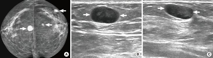
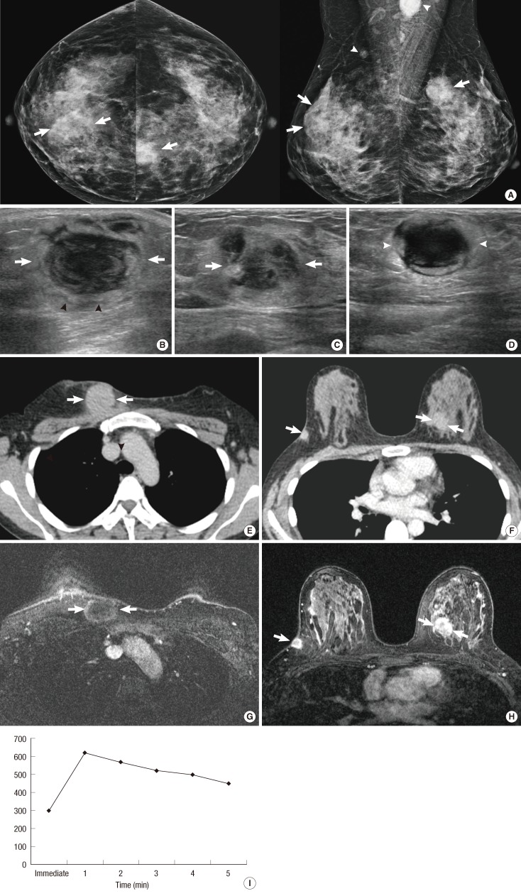
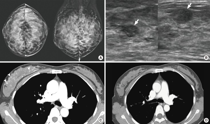
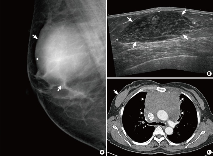
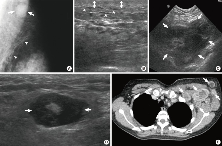
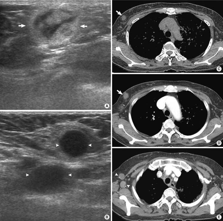
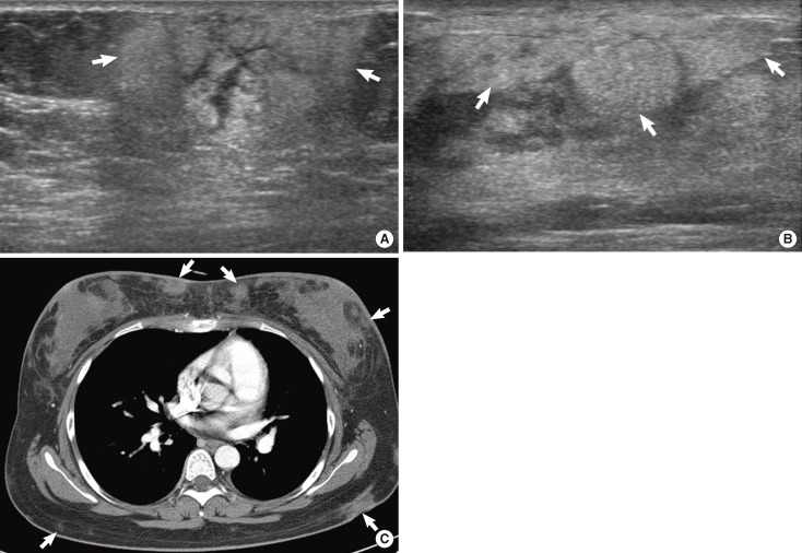
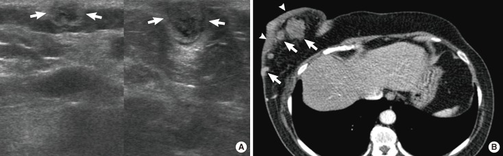
Hematological malignancies rarely affect the breast, and the majority of those that do are lymphomas. In this review, we describe the clinical aspects and multimodal imaging findings of breast lymphoma. We also illustrate the key clinical and radiological findings that allow it to be distinguished from various other malignant and benign diseases of the breast. Breast lymphoma manifests as a breast mass, a change in the subcutaneous tissue or the skin, or enlargement of the associated lymph node on radiological examination. Radiological findings associated with other breast malignancies, such as calcifications, spiculations, or architectural distortions are extremely rare. Skin and subcutaneous changes frequently accompany T-cell lymphoma. Multimodal breast imaging characteristics may aid in the diagnosis of breast lymphoma.

Keywords: Breast; Computed tomography; Lymphoma; Magnetic resonance imaging; Ultrasonography.


Conflict of interest statement

The authors declare that they have no competing interests.

Figures

Figure 1
Figure 1
A 61-year-old woman with diffuse large B-cell lymphoma. (A) Both craniocaudal mammograms show multiple circumscribed oval or round masses (arrows) in both breasts. On (B) right and (C) left breast ultrasonography, the masses (arrows) are circumscribed and very low echoic, mimicking cysts.
Figure 2
Figure 2
A 43-year-old woman with diffuse large B-cell lymphoma. (A) Both craniocaudal and mediolateral oblique mammograms demonstrate bilateral indistinct oval or round masses (arrows) and axillary lymph node enlargements (white arrowheads). (B) Ultrasonography (US) scan of the right breast shows an indistinct oval hypoechoic mass (arrows) with onion peel-like rims (black arrowheads). (C) US scan of the left breast shows an indistinct irregular hypoechoic mass (arrows). (D) US scan of the left axilla demonstrates an enlarged lymph node (white arrowheads). The node has indistinct margins and is very hypoechoic. (E, F) Enhanced breast computed tomography images show bilateral masses (arrows) with homogeneous enhancement. (G, H) Enhanced T1-weighted transverse magnetic resonance images show bilateral masses (arrows) with peripheral rim enhancement. (I) A time-enhancement curve obtained from the mass in left breast reveals rapid initial and washout delayed phase enhancement.
Figure 3
Figure 3
A 46-year-old woman with diffuse large B-cell lymphoma. (A) Both craniocaudal and mediolateral oblique mammograms are negative. (B) Ultrasonography scans of the right breast show multiple circumscribed oval hypoechoic masses (arrows). (C) Enhanced breast computed tomography (CT) image shows enlargement of right breast with multiple small enhancing foci (arrowheads). (D) Enhanced breast CT image obtained after chemotherapy shows the disappearance of enhancing foci in the right breast.
Figure 4
Figure 4
A 52-year-old woman with precursor T-lymphoblastic lymphoma. (A) Right mediolateral oblique mammogram shows a circumscribed and partially obscured marginated round hyperdense mass (arrows). (B) Ultrasonography scan of the right breast demonstrates a circumscribed oval hypoechoic mass (arrows). (C) Enhanced computed tomography scan demonstrates an oval isodense mass (arrow) in right breast and mediastinal widening (arrowheads) due to lymphadenopathy.
Figure 5
Figure 5
A 43-year-old woman with diffuse large B-cell lymphoma. (A) Left mediolateral oblique mammogram shows axillary lymphadenopathy (arrows) and trabecular thickening (arrowheads). (B) Ultrasonography (US) scans of the left breast show skin thickening (arrows) and dilated dermal lymphatics (arrowheads). (C, D) US scans of the left axilla show enlarged lymph nodes (arrows). One node is indistinct and irregular in shape (C) and the other is circumscribed and ovoid (D). The nodes have cortical thickening. (E) Enhanced breast computed tomography image shows multiple-rim enhancing lymph nodes (arrows) in left axilla.
Figure 6
Figure 6
A 65-year-old woman with diffuse large B-cell lymphoma. (A) Ultrasonography (US) scan of the right breast shows an indistinct oval hyperechoic mass (arrows) in the subcutaneous fat layer. (B) US scan of the right axilla shows enlarged lymph nodes with loss of internal fatty hila (arrowheads). (C) Pre-enhanced and (D) enhanced breast computed tomography (CT) images demonstrate an indistinct oval isodense mass (arrows) in right breast. The mass in right breast reveals 39 Hounsfield unit (HU) on pre-enhanced image and 45 HU on enhanced images. (E) Multiple enlarged axillary lymph nodes (arrowheads) are seen on enhanced CT scan.
Figure 7
Figure 7
A 21-year-old woman with NK/T-cell lymphoma. (A) Ultrasonography (US) scan of the right breast shows an indistinct oval hyperechoic mass (arrows) with internal tubular hypoechogenicities and thickening of overlying skin. (B) US scan of the left breast shows increased echotexture of subcutaneous fat layer (arrows). (C) Enhanced breast computed tomography scan shows multiple indistinct isodense masses (arrows) in both breasts and back, and diffuse skin thickening in both breasts.
Figure 8
Figure 8
A 62-year-old woman with peripheral T-cell lymphoma. (A) Ultrasonography (US) scans of the right breast show multiple oval or round masses (arrows) in skin and subcutaneous fat layers. (B) Enhanced computed tomography scan of right breast shows multiple isodense or hyperdense masses (arrows) with skin thickening (arrowheads).

References

    1. Swerdlow SH International Agency for Research on Cancer. WHO Classification of Tumours of Haematopoietic and Lymphoid Tissues. 4th ed. Lyon: IARC Press; 2008.
    1. Tavassoli FA, Devilee P International Agency for Research on Cancer. Pathology and Genetics of Tumours of the Breast and Female Genital Organs. Lyon: IARC Press; 2003. pp. 9–112.
    1. Giardini R, Piccolo C, Rilke F. Primary non-Hodgkin's lymphomas of the female breast. Cancer. 1992;69:725–735. - PubMed
    1. Dao AH, Adkins RB, Jr, Glick AD. Malignant lymphoma of the breast: a review of 13 cases. Am Surg. 1992;58:792–796. - PubMed
    1. Brogi E, Harris NL. Lymphomas of the breast: pathology and clinical behavior. Semin Oncol. 1999;26:357–364. - PubMed

LinkOut - more resources