

Using bosentan to treat paraquat poisoning-induced acute lung injury in rats
- PMID: 24155875
- PMCID: PMC3796527
- DOI: 10.1371/journal.pone.0075943
Using bosentan to treat paraquat poisoning-induced acute lung injury in rats
Abstract
Background: Paraquat poisoning is well known for causing multiple organ function failure (MODS) and high mortality. Acute lung injury and advanced pulmonary fibrosis are the most serious complications. Bosentan is a dual endothelin receptor antagonist. It plays an important role in treating PF. There is no related literature on the use of bosentan therapy for paraquat poisoning.
Objective: To study the use of bosentan to treat acute lung injury and pulmonary fibrosis as induced by paraquat.
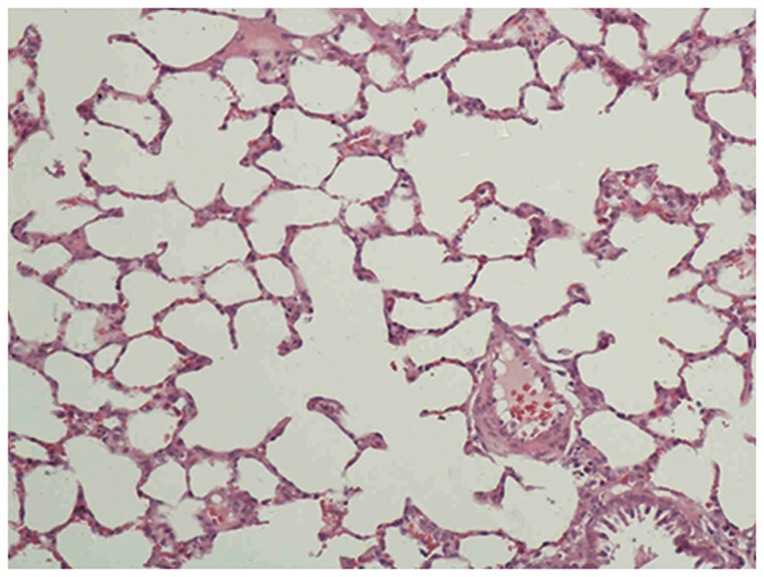
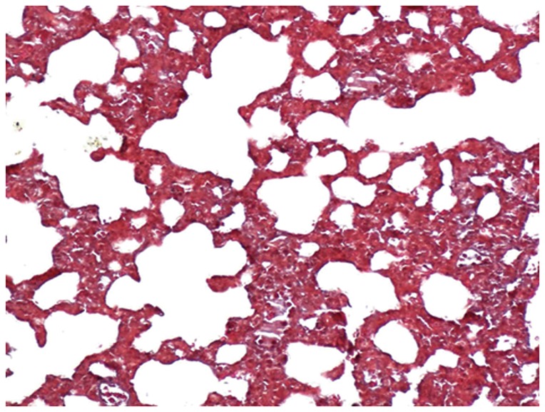
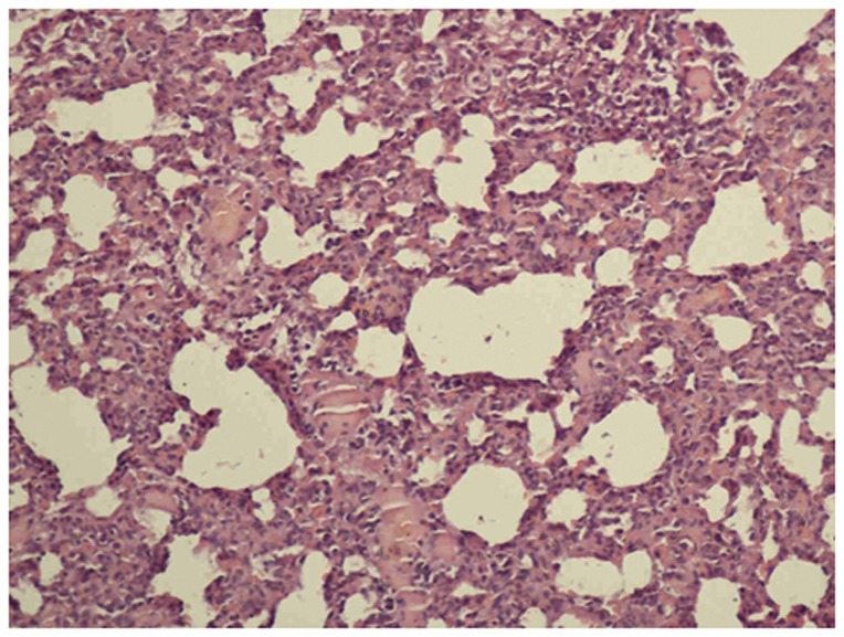
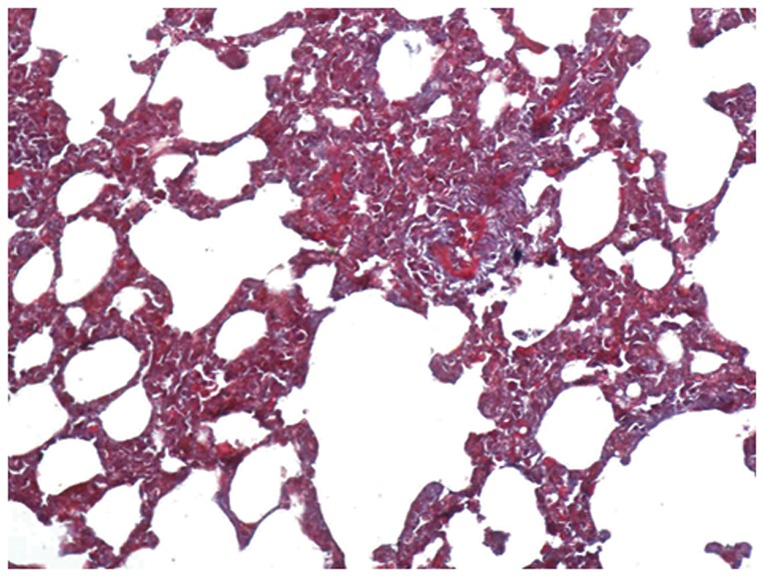
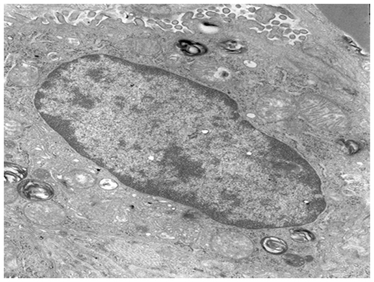
Method: A total of 120 adult Wister male rats were randomly assigned to three groups: the paraquat poisoning group (rats were intragastrically administered with paraquat at 50 mg/kg body weight once at the beginning); the bosentan therapy group (rats were administered bosentan at 100 mg/kg body weight by intragastric administration half an hour after paraquat was administered, then the same dose was administered once a day); and a control group (rats were administered intragastric physiological saline). On the 3rd, 7th, 14th, and 21st days following paraquat exposure, rats were sacrificed, and samples of lung tissue and venous blood were collected. The levels of transforming growth factor-β1 (TGF-β1), endothelin-1 (ET-1), and hydroxyproline (HYP) in the plasma and lung homogenate were determined. Optical and electronic microscopes were used to examine pathological changes.
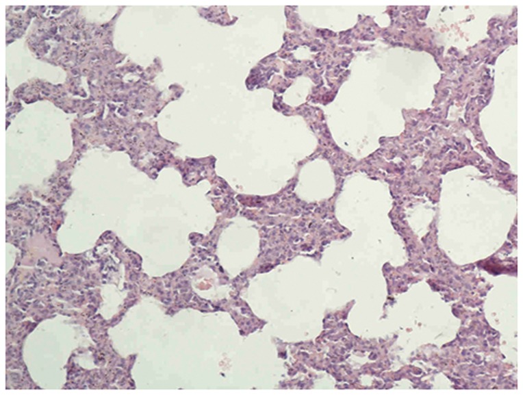
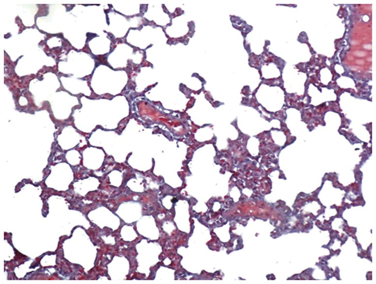
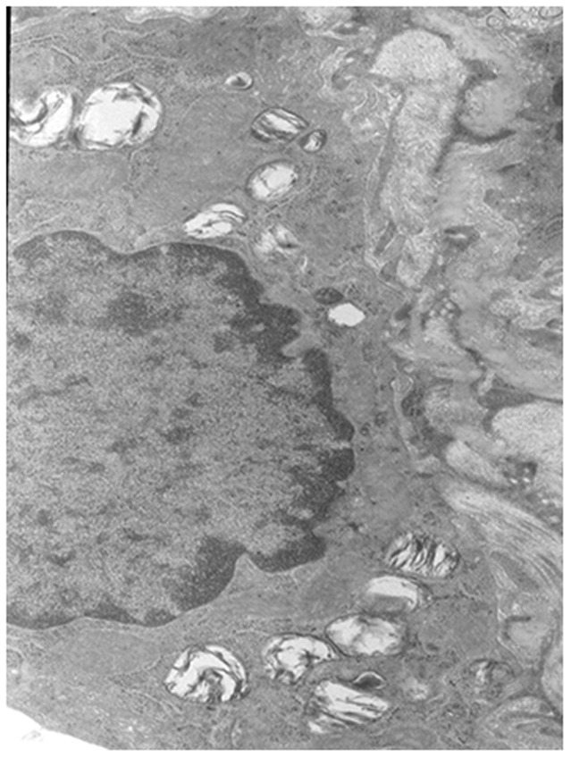
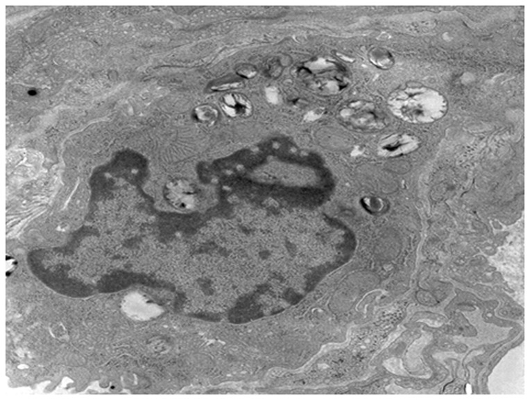
Result: The TGF-β1, ET-1, and HYP of the paraquat poisoning group were significantly higher than in the control group, and they were significantly lower in the 21st day therapy group than in the paraquat poisoning group on the same day. Under the optical and electronic microscopes, lung tissue damage was observed to be more severe but was then reduced after bosentan was administered.
Conclusion: Bosentan can reduce inflammation factor release. It has a therapeutic effect on acute lung injury as induced by paraquat.
Conflict of interest statement
Figures
References
-
- Chen HW, Tseng TK, Ding LW (2009) Intravenous paraquat poisoning. J Chin Med Assoc 72: 547–550. - PubMed
-
- Zhao B, Jia XD, Zhang ZC (2010) Relationship between paraquat tissue content and organ injury in paraquat poisoning rats. Zhonghua Lao Dong Wei Sheng Zhi Ye Bing Za Zhi 28: 220–223 [Article in Chinese]. - PubMed
-
- Choi Y, Cho K, Yoon S, Lee H, Choi Y (2008) A case of paraquat intoxication caused by intravenous injection. Am J Emerg Med 26: 836.e3–4. - PubMed
-
- Mohammadi-Karakani A, Ghazi-Khansari M, Sotoudeh M (2006) Lisinopril ameliorates paraquat-induced lung fibrosis. Clin Chim Acta 367: 170–174. - PubMed
Publication types
MeSH terms
Substances
LinkOut - more resources
Full Text Sources
Other Literature Sources
