. 2013 Oct 14;8(10):e77771.
doi: 10.1371/journal.pone.0077771. eCollection 2013.

Improved PCR performance using template DNA from formalin-fixed and paraffin-embedded tissues by overcoming PCR inhibition

Affiliations

Improved PCR performance using template DNA from formalin-fixed and paraffin-embedded tissues by overcoming PCR inhibition

Dimo Dietrich et al. PLoS One. .

Abstract

Formalin-fixed and paraffin-embedded (FFPE) tissues represent a valuable source for biomarker studies and clinical routine diagnostics. However, they suffer from degradation of nucleic acids due to the fixation process. Since genetic and epigenetic studies usually require PCR amplification, this degradation hampers its use significantly, impairing PCR robustness or necessitating short amplicons. In routine laboratory medicine a highly robust PCR performance is mandatory for the clinical utility of genetic and epigenetic biomarkers. Therefore, methods to improve PCR performance using DNA from FFPE tissue are highly desired and of wider interest. The effect of template DNA derived from FFPE tissues on PCR performance was investigated by means of qPCR and conventional PCR using PCR fragments of different sizes. DNA fragmentation was analyzed via agarose gel electrophoresis. This study showed that poor PCR amplification was partly caused by inhibition of the DNA polymerase by fragmented DNA from FFPE tissue and not only due to the absence of intact template molecules of sufficient integrity. This PCR inhibition was successfully minimized by increasing the polymerase concentration, dNTP concentration and PCR elongation time thereby allowing for the robust amplification of larger amplicons. This was shown for genomic template DNA as well as for bisulfite-converted template DNA required for DNA methylation analyses. In conclusion, PCR using DNA from FFPE tissue suffers from inhibition which can be alleviated by adaptation of the PCR conditions, therefore allowing for a significant improvement of PCR performance with regard to variability and the generation of larger amplicons. The presented solutions to overcome this PCR inhibition are of tremendous value for clinical chemistry and laboratory medicine.


Conflict of interest statement

Competing Interests: The authors have declared that no competing interests exist.

Figures

Figure 1
Figure 1. Degradation of DNA from FFPE Tissues and its Effect on PCR Amplification of Amplicons with Different Sizes.
Analysis of DNA integrity of genomic and bisulfite-converted DNA from unfixed and FFPE tissues by means of (A) agarose gel electrophoresis and (B) end-point PCR using PCR fragments of different sizes within the PITX2 gene locus. (C) qPCR results applying increasing amounts (2.5–3,840 ng) of genomic template DNA from unfixed and (D) from FFPE tissue. Shown are the mean values (± standard deviations) from triplicate measurements. Each PCR was performed with 1 U Taq polymerase. DNA from unfixed specimens is considered high molecular weight (HMW) DNA.
Figure 2
Figure 2. Inhibitory Effect of Template DNA from FFPE Tissues on PCR Performance.
qPCR with 1 µg of genomic HMW template DNA and increasing amounts (60-1,440 ng) of spiked genomic template DNA from FFPE tissue Genomic DNA from FFPE tissue was treated beforehand with active DNase I (+) and heat-inactivated DNase I (-), respectively. qPCR was performed using a 150-bp fragment and 1 U Taq polymerase. Shown are the mean values (± standard deviations) from triplicate measurements.
Figure 3
Figure 3. Reduction of PCR Inhibition by Increased Amounts of Taq Polymerase.
qPCR with HMW and template DNA from FFPE tissue using different amounts of Taq polymerase. PCR-amplification of increasing amounts of genomic HMW template DNA (2.5-3,840 ng) using (A) 2 U Taq, (B) 4 U Taq and PCR-amplification of template DNA from FFPE tissue using (C) 2 U Taq, (D) 4 U Taq. Shown are the mean values (± standard deviations) from triplicate measurements.
Figure 4
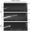
Figure 4. Successful PCR Amplification of Larger Fragments by Overcoming PCR Inhibition.
PCR-amplified DNA fragments of different sizes within the PITX2 gene locus using template DNA from FFPE tissue. The PCR was carried out using 1 µg (upper and middle panel) and 5 ng (lower panel) template DNA in the presence of 1 U and 4 U Taq DNA polymerase, respectively.
Figure 5
Figure 5. Transferability of the Findings to a qPCR Targeting an Alternative Genomic Locus.
qPCR applying a 200-bp PCR fragment within the ACTB gene locus. Amplification with (A) HMW DNA from unfixed tissue and (B) DNA from FFPE tissue (2.5 ng to 3,840 ng) in the presence of 1 U and 4 U Taq DNA polymerase. Shown are the mean values (± standard deviations) from triplicate measurements.
Figure 6
Figure 6. PCR Inhibition by Template DNA from FFPE Tissues with Regard to PCR Conditions (dNTP Concentration and Thermal Cycling Profile).
Shown are qPCR results applying a 200-bp fragment within the PITX2 gene locus using template DNA from FFPE tissue (80-1,920 ng) in the presence of 1 U Taq polymerase. (A) dNTP concentrations and (B) annealing and elongation times were varied. Shown are the mean values (± standard deviations) from triplicate measurements.
Figure 7
Figure 7. Inhibitory Effect of Template DNA from FFPE Tissues on PCR with Pfu Polymerase.
A 75-bp PCR fragment within the PITX2 gene locus was amplified using genomic template DNA from FFPE tissue (placenta) in the presence of 1 U and 2 U Pfu polymerase. Shown are the mean values (± standard deviations) from triplicate measurements.
Figure 8
Figure 8. PCR Inhibition by Bisulfite-Converted Template DNA from FFPE Tissues.
(A) qPCR–amplification of different amounts (10–3,840 ng) of bisulfite-converted template DNA from FFPE tissue using a 129-bp PCR fragment within the ACTB gene locus. 1 U and 4 U Taq polymerase were used for qPCR. Shown are the mean values (± standard deviations) from triplicate measurements. (B) PCR amplification of the specific PCR product was confirmed by agarose gel electrophoresis using 1 U (upper panel) and 4 U (lower panel) Taq polymerase.

References

    1. Blow N (2007) Tissue preparation: Tissue issues. Nature 448: 959-963. doi:10.1038/448959b. PubMed: 17713539. - DOI - PubMed
    1. Bereczki L, Kis G, Bagdi E, Krenacs L (2007) Optimization of PCR amplification for B- and T-cell clonality analysis on formalin-fixed and paraffin-embedded samples. Pathol Oncol Res 13: 209-214. doi:10.1007/BF02893501. PubMed: 17922050. - DOI - PubMed
    1. Kuykendall JR, Bogdanffy MS (1992) Efficiency of DNA-histone crosslinking induced by saturated and unsaturated aldehydes in vitro. Mutat Res 283: 131-136. doi:10.1016/0165-7992(92)90145-8. PubMed: 1381490. - DOI - PubMed
    1. Bonin S, Petrera F, Niccolini B, Stanta G (2003) PCR analysis in archival postmortem tissues. Mol Pathol 56: 184-186. doi:10.1136/mp.56.3.184. PubMed: 12782767. - DOI - PMC - PubMed
    1. Gilbert MT, Haselkorn T, Bunce M, Sanchez JJ, Lucas SB et al. (2007) The isolation of nucleic acids from fixed, paraffin-embedded tissues-which methods are useful when? PLOS ONE 2: e537. doi:10.1371/journal.pone.0000537. PubMed: 17579711. - DOI - PMC - PubMed