. 2013 Oct 14;8(10):e77843.
doi: 10.1371/journal.pone.0077843. eCollection 2013.

Altered cardiac electrophysiology and SUDEP in a model of Dravet syndrome

Affiliations

Altered cardiac electrophysiology and SUDEP in a model of Dravet syndrome

David S Auerbach et al. PLoS One. .

Abstract

Objective: Dravet syndrome is a severe form of intractable pediatric epilepsy with a high incidence of SUDEP: Sudden Unexpected Death in epilepsy. Cardiac arrhythmias are a proposed cause for some cases of SUDEP, yet the susceptibility and potential mechanism of arrhythmogenesis in Dravet syndrome remain unknown. The majority of Dravet syndrome patients have de novo mutations in SCN1A, resulting in haploinsufficiency. We propose that, in addition to neuronal hyperexcitability, SCN1A haploinsufficiency alters cardiac electrical function and produces arrhythmias, providing a potential mechanism for SUDEP.

Methods: Postnatal day 15-21 heterozygous SCN1A-R1407X knock-in mice, expressing a human Dravet syndrome mutation, were used to investigate a possible cardiac phenotype. A combination of single cell electrophysiology and in vivo electrocardiogram (ECG) recordings were performed.

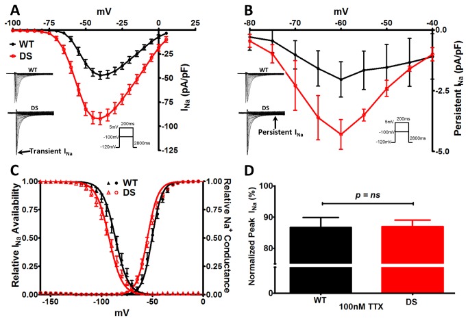
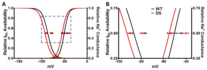
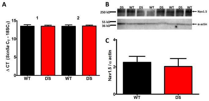
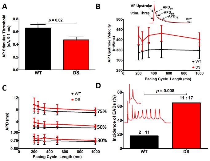
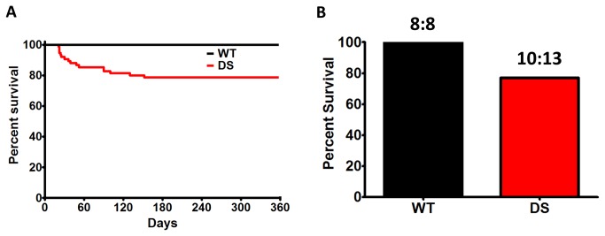
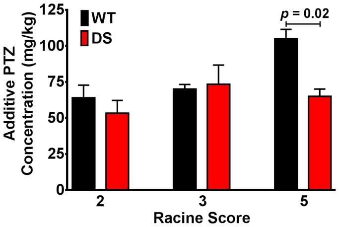
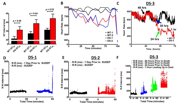
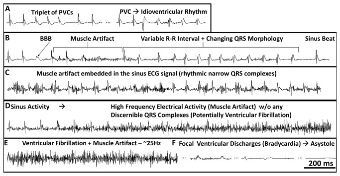
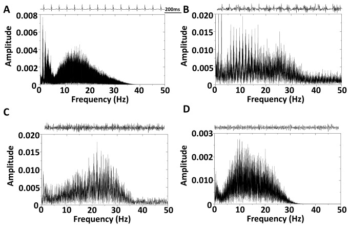
Results: We observed a 2-fold increase in both transient and persistent Na(+) current density in isolated Dravet syndrome ventricular myocytes that resulted from increased activity of a tetrodotoxin-resistant Na(+) current, likely Nav1.5. Dravet syndrome myocytes exhibited increased excitability, action potential duration prolongation, and triggered activity. Continuous radiotelemetric ECG recordings showed QT prolongation, ventricular ectopic foci, idioventricular rhythms, beat-to-beat variability, ventricular fibrillation, and focal bradycardia. Spontaneous deaths were recorded in 2 DS mice, and a third became moribund and required euthanasia.

Interpretation: These data from single cell and whole animal experiments suggest that altered cardiac electrical function in Dravet syndrome may contribute to the susceptibility for arrhythmogenesis and SUDEP. These mechanistic insights may lead to critical risk assessment and intervention in human patients.


Conflict of interest statement

Competing Interests: The authors have declared that no competing interests exist.

Figures

Figure 1
Figure 1. DS Mice Have Altered Cardiac INa Properties.
A. Current-voltage (I-V) relationship of transient INa. Peak transient INa density is increased 2-fold in the DS (N = 6, n = 14) vs WT cardiac myocytes (N = 8, n = 20, p < 0.0001). Inset: Representative traces from each group. B. I-V relationship for persistent INa (pre- minus post-30 µM TTX) also shows a 2-fold increase in peak persistent INa in the DS vs. WT groups. To further confirm these results we employed the P/4 method to measure the persistent INa, yielding similar results (-60 mV, WT, -1.72 ± 0.50; DS, -3.88 ± 0.72, N = 2, n = 5-9, p = 0.02). C. Leftward shift (V½ of Boltzman fit, p = 0.04) in the voltage dependence of INa availability and conductance in the DS group. D. Similar percent change in peak transient INa density upon administration of 100 nM TTX in the WT and DS groups. Unpaired t-test with Welch’s correction.
Figure 2
Figure 2. Isolation of TTX-R and TTX-S INa Biophysical Properties.
A. Boltzman curves for the voltage dependence of INa availability and conductance for the total cardiac INa (TTX-S + TTX-R INa; reproduction of the curve-fits from Figure 1C). In both WT and DS myocytes the V½ values of TTX-R INa (closed circles, following blockade of TTX-S INa with 100 nM TTX) and TTX-S INa (open circles, defined as total INa minus TTX-R INa) are plotted. Pharmacological separation of TTX-S and TTX-R INa was confirmed by the loss of difference in the V½ values between WT vs DS, and the development of a significant difference between the TTX-S vs. TTX-R V ½ values for INa availability and conductance. B. Zoom-in of the boxed region in A.
Figure 3
Figure 3. mScn5a and Nav1.5 levels are unchanged in DS mutant hearts.
A. Heart RNA from biological replicates (DS mice, n = 4; WT mice, n = 5) were used to generate two independent cDNAs per animal. The cDNAs were assayed using qPCR in quadruplicate with two independent Scn5a TaqMan primer sets and normalized to 18s RNA. B. Western blots of membrane proteins isolated from DS and WT ventricular CMs. 50 µg of protein was loaded in each lane, and probed with anti-Nav1.5 (Mohler 1:1000), and anti-α-actin (Sigma 1:500), which served as the loading control. C. Quantification of Nav1.5 expression normalized to α-actin expression.
Figure 4
Figure 4. DS myocytes exhibit increased excitability and incidence of early after depolarizations (EADs).
A. DS myocytes require significantly less injected current to fire APs. B. DS myocyte AP upstroke velocity is faster at all pacing cycle lengths (p = ns). C. Slight prolongation of the AP duration at 30%, 50%, and 75% repolarization at many pacing cycle lengths (p = ns). D. DS myocytes are significantly more susceptible to EADs, a substrate for arrhythmogenesis. Inset: Representative EADs from DS myocytes (red.) Panels A-C, unpaired t-test with Welch’s correction. Panel D χ2 Test (WT, N = 9, n = 11, DS, N = 8, n = 17).
Figure 5
Figure 5. DS Mice Undergo SUDEP.
A. Kaplan-Meier survival curves for WT and DS mice (N = 75 for each group, p < 0.0001, Log-rank, Mantel-Cox, Survival Test). B. Percent survival in WT (N = 8) and DS (N = 13) mice implanted with radiotelemetry units. SUDEP or near-SUDEP in 3 DS mice (at P41, P45, and P51, respectively).
Figure 6
Figure 6. Decreased Threshold for PTZ Induced Seizures in DS Mice.
WT and DS mice were administered incremental doses of pentylenetetrazole (PTZ), monitored for observable seizures, and classified on the Racine Scale.
Figure 7
Figure 7. Altered Heart Rates Precede Death.
A. DS mice exhibit significant QT prolongation (50 - 90%). B. Heart rates in DS mice decrease 100 min before death, followed by a sharp increase just prior to the terminal event, while the WT heart rates remains high and constant. (100 minutes = 10:16 PM in WT-1 and DS-1; 7:46 PM in WT-2 and DS-2). C. WT-3 and DS-3 HR cycling, followed by DS exhibiting sudden drops in heart rate in the 72 h preceding death. D and E. Increased R-R variability 60 min prior to SUDEP in DS-1 (blue) and DS-2 (red), respectively, with further increased variability immediately preceding the lethal arrhythmia, while 1 day prior at the same time the R-R interval was constant (black). F. Progressive bradycardia and increased R-R variability in DS-3 at several time points preceding an agonal state and euthanasia (denoted by colored arrows in C).
Figure 8
Figure 8. Cardiac Arrhythmias Precede SUDEP in DS.
Lead II ECG traces illustrating cardiac arrhythmias preceding death. A-C. In mouse DS-2, muscle artifact consistent with convulsive seizures was preceded by idioventricular rhythms, including premature ventricular complexes (PVCs), bundle branch block (BBB), altered QRS morphology, and R-R variability. D and E. Initiation of high frequency electrical activity without any discernible sinus activity, consistent with VF. F. Low amplitude wide complex focal bradycardia with a BBB morphology, and eventual asystole.
Figure 9
Figure 9. Dominant Frequency Analysis.
A. Sinus rhythm 1 day prior to SUDEP, which is consistent with heart rate (728 bpm) analysis. B. Muscle artifact embedded in the sinus ECG (same as Figure 9 C) without any clear frequency peaks. C. High frequency electrical activity without any discernible sinus activity, consistent with VF (~25 Hz, same as Figure 9, D and E). D. PTZ induced seizures lead to a lower frequency electrical signal (~10 - 20 Hz). Inset: Representative snapshots of the ECG signal included in the fast-fourier transformation.

References

    1. Escayg A, Goldin AL (2010) Sodium channel SCN1A and epilepsy: mutations and mechanisms. Epilepsia 51: 1650-1658. doi:10.1111/j.1528-1167.2010.02640.x. PubMed: 20831750. - DOI - PMC - PubMed
    1. Dravet C, Bureau M, Oguni H, Fukuyama Y, Cokar O (2005) Severe myoclonic epilepsy in infancy: Dravet syndrome. Adv_Neurol 95: 71-102. PubMed: 15508915. - PubMed
    1. Oakley JC, Kalume F, Catterall WA (2011) Insights into pathophysiology and therapy from a mouse model of Dravet syndrome. Epilepsia 52 Suppl 2: 59-61. doi:10.1111/j.1528-1167.2011.03004.x. PubMed: 21463282. - DOI - PMC - PubMed
    1. Surges R, Adjei P, Kallis C, Erhuero J, Scott CA et al. (2010) Pathologic cardiac repolarization in pharmacoresistant epilepsy and its potential role in sudden unexpected death in epilepsy: a case-control study. Epilepsia 51: 233-242. doi:10.1111/j.1528-1167.2009.02330.x. PubMed: 19817816. - DOI - PubMed
    1. Shorvon S, Tomson T (2011) Sudden unexpected death in epilepsy. Lancet, 378: 2028–38. PubMed: 21737136. - PubMed

Publication types

MeSH terms

LinkOut - more resources