

Interactions of Candida albicans with host epithelial surfaces
- PMID: 24155995
- PMCID: PMC3805843
- DOI: 10.3402/jom.v5i0.22434
Interactions of Candida albicans with host epithelial surfaces
Abstract
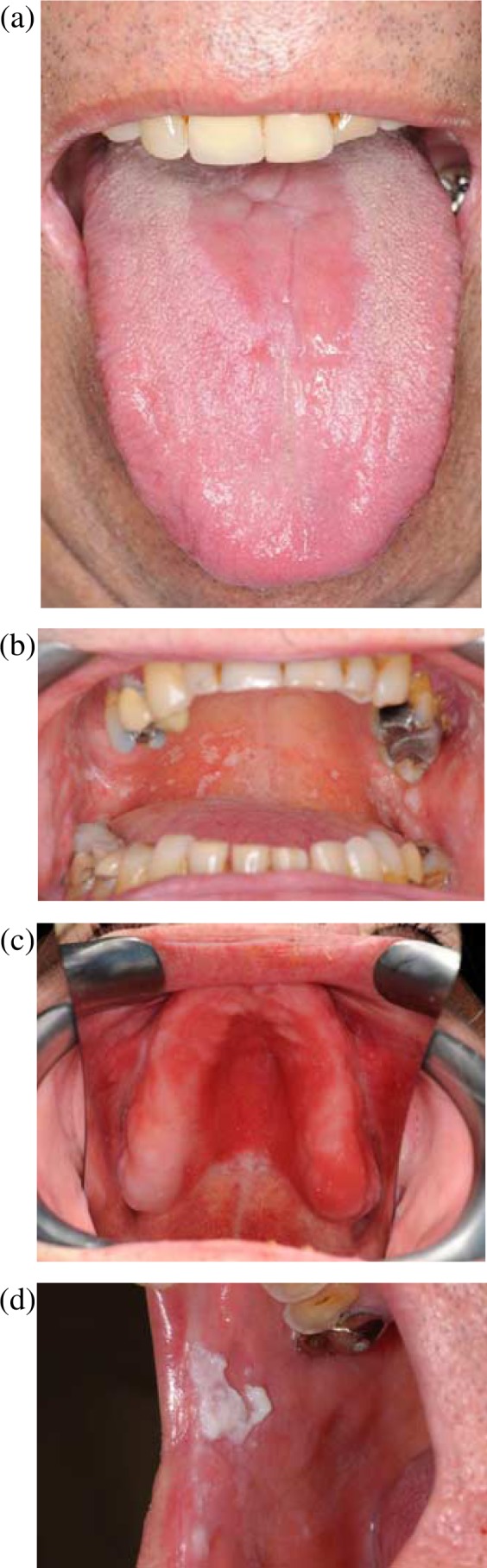
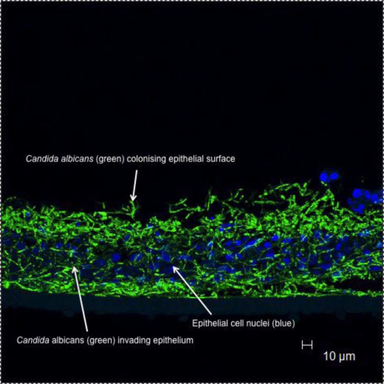
Candida albicans is an opportunistic, fungal pathogen of humans that frequently causes superficial infections of oral and vaginal mucosal surfaces of debilitated and susceptible individuals. The organism is however, commonly encountered as a commensal in healthy individuals where it is a component of the normal microflora. The key determinant in the type of relationship that Candida has with its host is how it interacts with the epithelial surface it colonises. A delicate balance clearly exists between the potentially damaging effects of Candida virulence factors and the nature of the immune response elicited by the host. Frequently, it is changes in host factors that lead to Candida seemingly changing from a commensal to pathogenic existence. However, given the often reported heterogeneity in morphological and biochemical factors that exist between Candida species and indeed strains of C. albicans, it may also be the fact that colonising strains differ in the way they exploit resources to allow persistence at mucosal surfaces and as a consequence this too may affect the way Candida interacts with epithelial cells. The aim of this review is to provide an overview of some of the possible interactions that may occur between C. albicans and host epithelial surfaces that may in turn dictate whether Candida removal, its commensal persistence or infection follows.
Keywords: biofilm; oral microbiology; pathogenesis; virulence factors.
Figures
References
-
- Leli C, Mencacci A, Meucci M, Bietolini C, Vitali M, Farinelli S, et al. Association of pregnancy and Candida vaginal colonization in women with or without symptoms of vulvovaginitis. Minerva Ginecol. 2013;65:303–9. - PubMed
-
- Foxman B, Marsh JV, Gillespie B, Sobel JD. Frequency and response to vaginal symptoms among white and African American women: results of a random digit dialing survey. J Womens Health. 1998;7:1167–74. - PubMed
Publication types
LinkOut - more resources
Full Text Sources
Other Literature Sources