

Achromatic miniature lens system for coherent Raman scattering microscopy
- PMID: 24156075
- PMCID: PMC3799677
- DOI: 10.1364/BOE.4.002196
Achromatic miniature lens system for coherent Raman scattering microscopy
Abstract
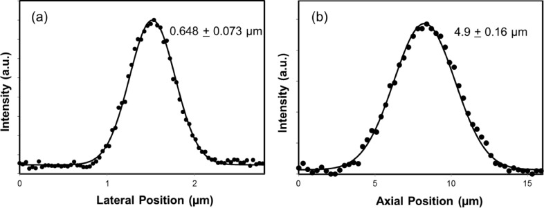
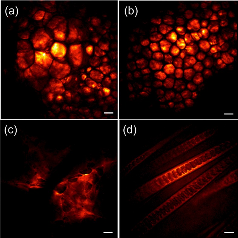
We discuss the design and performance of a miniature objective lens optimized for coherent Raman scattering microscopy. The packaged lens assembly has a numerical aperture of 0.51 in water and an outer diameter of 8 mm. The lens system exhibits minimum chromatic aberrations, and produces coherent Raman scattering images with sub-micrometer lateral resolution (0.648 μm) using near-infrared excitation pulses. We demonstrate that despite the small dimensions of the miniature objective, the performance of this lens system is comparable to standard microscope objective lenses, offering opportunities for miniaturizing coherent Raman scattering imaging probes without sacrificing the image quality.
Keywords: (180.4315) Nonlinear microscopy; (220.3630) Lenses; (350.3950) Micro-optics.
Figures


Similar articles
-
Coherent Raman scattering imaging with a near-infrared achromatic metalens.APL Photonics. 2021 Sep 1;6(9):096107. doi: 10.1063/5.0059874. Epub 2021 Sep 14. APL Photonics. 2021. PMID: 34553044 Free PMC article.
-
Increasing the imaging depth of coherent anti-Stokes Raman scattering microscopy with a miniature microscope objective.Opt Lett. 2007 Aug 1;32(15):2212-4. doi: 10.1364/ol.32.002212. Opt Lett. 2007. PMID: 17671587
-
High-resolution multimodal flexible coherent Raman endoscope.Light Sci Appl. 2018 May 30;7:10. doi: 10.1038/s41377-018-0003-3. eCollection 2018. Light Sci Appl. 2018. PMID: 30839624 Free PMC article.
-
Contributed Review: A new synchronized source solution for coherent Raman scattering microscopy.Rev Sci Instrum. 2016 Jul;87(7):071501. doi: 10.1063/1.4955474. Rev Sci Instrum. 2016. PMID: 27475540 Review.
-
Coherent anti-stokes Raman scattering microscopy for high speed non- staining biomolecular imaging.Curr Pharm Biotechnol. 2013;14(2):150-8. Curr Pharm Biotechnol. 2013. PMID: 22356111 Review.
Cited by
-
Coherent Raman scattering imaging with a near-infrared achromatic metalens.APL Photonics. 2021 Sep 1;6(9):096107. doi: 10.1063/5.0059874. Epub 2021 Sep 14. APL Photonics. 2021. PMID: 34553044 Free PMC article.
-
Introduction: feature issue on optical molecular probes, imaging, and drug delivery.Biomed Opt Express. 2014 Jan 29;5(2):643-4. doi: 10.1364/BOE.5.000643. eCollection 2014 Feb 1. Biomed Opt Express. 2014. PMID: 24575356 Free PMC article.
-
Label-free Imaging of Myocardial Remodeling in Atrial Fibrillation Using Nonlinear Optical Microscopy: A Feasibility Study.J Atr Fibrillation. 2018 Feb 28;10(5):1644. doi: 10.4022/jafib.1644. eCollection 2018 Feb. J Atr Fibrillation. 2018. PMID: 29988238 Free PMC article.
References
-
- Cheng J. X., Xie X. S., Eds., Coherent Raman Scattering Microscopy (CRC Press, 2013).
-
- Henry F. P., Côté D., Randolph M. A., Rust E. A. Z., Redmond R. W., Kochevar I. E., Lin C. P., Winograd J. M., “Real-time in vivo assessment of the nerve microenvironment with coherent antiStokes Raman scattering microscopy,” Plastic and reconstructive surgery 123, 123S–130S (2009).10.1097/PRS.0b013e318191c5b8 - DOI - PubMed
Grants and funding
LinkOut - more resources
Full Text Sources
Other Literature Sources