. 2013 Oct 25;10(1):55.
doi: 10.1186/1743-8977-10-55.

Predictive value of in vitro assays depends on the mechanism of toxicity of metal oxide nanoparticles

Affiliations

Predictive value of in vitro assays depends on the mechanism of toxicity of metal oxide nanoparticles

Wan-Seob Cho et al. Part Fibre Toxicol. .

Abstract

Background: Hazard identification for risk assessment of nanoparticles (NPs) is mainly composed of in vitro cell-based assays and in vivo animal experimentation. The rapidly increasing number and functionalizations of NPs makes in vivo toxicity tests undesirable on both ethical and financial grounds, creating an urgent need for development of in vitro cell-based assays that accurately predict in vivo toxicity and facilitate safe nanotechnology.

Methods: In this study, we used 9 different NPs (CeO2, TiO2, carbon black, SiO2, NiO, Co3O4, Cr2O3, CuO, and ZnO). As an in vivo toxicity endpoint, the acute lung inflammogenicity in a rat instillation model was compared with the in vitro toxicity endpoints comprising cytotoxicity, pro-inflammatory cytokine expression, or haemolytic potential. For in vitro assays, 8 different cell-based assays were used including epithelial cells, monocytic/macrophage cells, human erythrocytes, and combined culture.

Results: ZnO and CuO NPs acting via soluble toxic ions showed positive results in most of assays and were consistent with the lung inflammation data. When compared in in vitro assays at the same surface area dose (30 cm2/mL), NPs that were low solubility and therefore acting via surface reactivity had no convincing activity, except for CeO2 NP. Cytotoxicity in differentiated peripheral blood mononuclear cells was the most accurate showing 89% accuracy and 11% false negativity in predicting acute lung inflammogenicity. However, the haemolysis assay showed 100% consistency with the lung inflammation if any dose, having statistical significance was considered positivity. Other cell-based in vitro assays showed a poorer correlation with in vivo inflammogenicity.

Conclusions: Based on the toxicity mechanisms of NPs, two different approaches can be applied for prediction of in vivo lung inflammogenicity. Most in vitro assays were good at detecting NPs that act via soluble ions (i.e., ZnO and CuO NP). However, in vitro assays were limited in detecting NPs acting via surface reactivity as their mechanism of toxicity, except for the haemolysis assay.


Figures
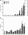
Figure 1
Figure 1
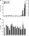
Cytotoxicity and IL-8 expression of A549 cells after exposure to NPs for 24 h. (A) Cytotoxicity was measured by trypan blue exclusion for ZnO and CuO NP whilst others were measured by LDH. (B) Levels of IL-8 of A549 cells at 24 h following treatment. Note that the surface area doses were 30, 100, and 300 cm2/mL except for ZnO and CuO NP which were 3, 10, and 30 cm2/mL. Values are mean ± SD from minimum four independent experiments. Significance versus vehicle control (VEH): *p < 0.05, ***p < 0.001.
Figure 2
Figure 2
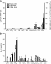
Cytotoxicity and IL-8 expression of 16-HBE cells after exposure to NPs for 24 h. (A) Cytotoxicity was measured by trypan blue exclusion for ZnO and CuO NP whilst others were measured by LDH. (B) Levels of IL-8 of 16-HBE cells at 24 h following treatment. Note that the surface area doses were 30, 100, and 300 cm2/mL except for ZnO and CuO NP which were 3, 10, and 30 cm2/mL. Values are mean ± SD from minimum four independent experiments. Significance versus vehicle control (VEH): *p < 0.05, ***p < 0.001.
Figure 3
Figure 3
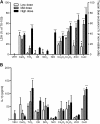
Cytotoxicity and IL-1β expression of monocytic THP-1 cells after exposure to NPs for 24 h. (A) Cytotoxicity was measured by trypan blue exclusion for ZnO and CuO NP whilst others were measured by LDH. (B) Levels of IL-1β of monocytic THP-1 cells at 24 h following treatment. Note that the surface area doses were 30, 100, and 300 cm2/mL except for ZnO and CuO NP which were 3, 10, and 30 cm2/mL. Values are mean ± SD from minimum four independent experiments. Significance versus vehicle control (VEH): *p < 0.05, **p < 0.01, ***p < 0.001.
Figure 4
Figure 4
Cytotoxicity and IL-1β expression of primary cultured alveolar macrophages after exposure to NPs for 24 h. (A) Cytotoxicity was measured by trypan blue exclusion for ZnO and CuO NP whilst others were measured by LDH. (B) Levels of IL-1β of primary cultured alveolar macrophages at 24 h following treatment. Note that the surface area doses were 30, 100, and 300 cm2/mL except for ZnO and CuO NP which were 3, 10, and 30 cm2/mL. Values are mean ± SD from minimum four independent experiments. Significance versus vehicle control (VEH): *p < 0.05, **p < 0.01, ***p < 0.001.
Figure 5
Figure 5
Cytotoxicity and pro-inflammatory cytokine expression of differentiated PBMC after exposure to NPs for 24 h. (A) Cytotoxicity was measured by trypan blue exclusion for ZnO and CuO NP whilst others were measured by LDH. (B) Levels of IL-1β; (C) levels of TNF-α. PBMC were differentiated by 5-day incubation. Note that the surface area doses were 30, 100, and 300 cm2/mL except for ZnO and CuO NP which were 3, 10, and 30 cm2/mL. Values are mean ± SD from minimum four independent experiments. Significance versus vehicle control (VEH): *p < 0.05, **p < 0.01, ***p < 0.001.
Figure 6
Figure 6
Cytotoxicity and pro-inflammatory cytokine expression of differentiated THP-1 cells after exposure to NPs for 24 h. (A) Cytotoxicity was measured by trypan blue exclusion for ZnO and CuO NP whilst others were measured by LDH. (B) Levels of IL-1β; (C) levels of TNF-α. (D) Treatment of NP with cytochalasin D (0.2 μM) showed marked decrease of IL-1β expression compared to NP without cytochalasin D (B). THP-1 cells were differentiated by treatment with PMA (10 ng/mL) and NPs were treated at 30, 100, and 300 cm2/mL except for ZnO NP (3, 10, and 30 cm2/mL) and CuO NP (0.3, 1, and 3 cm2/mL). Values are mean ± SD from minimum four independent experiments. Significance versus vehicle control (VEH): *p < 0.05, **p < 0.01, ***p < 0.001.
Figure 7
Figure 7
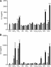
IL-8 expression by NP-conditioned media. A549 cells were exposed to conditioned media from NP-exposed differentiated THP-1 cells for 4 h. THP-1 cells were differentiated by treatment for PMA (10 ng/mL) and NPs were treated at surface area doses of 30, 100, and 300 cm2/mL except for ZnO NP (3, 10, and 30 cm2/mL) and CuO NP (0.3, 1, and 3 cm2/mL). (A) The levels of IL-8 in the conditioned media from THP-1 cells before addition of A549 cells. (B) The levels of IL-8 after addition of NP-free conditioned media to A549 cells. Values are mean ± SD from minimum four independent experiments. Significance versus vehicle control (VEH): ***p < 0.001.
Figure 8
Figure 8
Haemolysis assay of NP. NPs were treated at surface area doses of 30, 100, and 300 cm2/mL. Values are mean ± SD from minimum four independent experiments. Significance versus vehicle control (VEH): *p < 0.05, ***p < 0.001.

References

    1. Nel AE, Madler L, Velegol D, Xia T, Hoek EM, Somasundaran P, Klaessig F, Castranova V, Thompson M. Understanding biophysicochemical interactions at the nano-bio interface. Nat Mater. 2009;10:543–557. doi: 10.1038/nmat2442. - DOI - PubMed
    1. Borm PJ, Robbins D, Haubold S, Kuhlbusch T, Fissan H, Donaldson K, Schins R, Stone V, Kreyling W, Lademann J. et al.The potential risks of nanomaterials: a review carried out for ECETOC. Part Fibre Toxicol. 2006;10:11. doi: 10.1186/1743-8977-3-11. - DOI - PMC - PubMed
    1. Organisation for Economic Co-operation and Development. Nanosafety at the OECD: the first five years 2006–2010. Paris: OECD; 2011.
    1. Organisation for Economic Co-operation and Development. Six years of OECD work on the safety of manufactured nanomaterials: achievements and future opportunities. Paris: OECD; 2013.
    1. Donaldson K, Borm PJ, Castranova V, Gulumian M. The limits of testing particle-mediated oxidative stress in vitro in predicting diverse pathologies; relevance for testing of nanoparticles. Part Fibre Toxicol. 2009;10:13. doi: 10.1186/1743-8977-6-13. - DOI - PMC - PubMed

Publication types