. 2013 Oct 24:10:111.
doi: 10.1186/1742-4690-10-111.

90K, an interferon-stimulated gene product, reduces the infectivity of HIV-1

Affiliations

90K, an interferon-stimulated gene product, reduces the infectivity of HIV-1

Veronika Lodermeyer et al. Retrovirology. .

Abstract

Background: In response to viral infections, interferons induce the transcription of several hundred genes in mammalian cells. Specific antiviral functions, however, have only been attributed to a few of them. 90K/LGALS3BP has been reported to be an interferon-stimulated gene that is upregulated in individuals with cancer or HIV-1 infection.

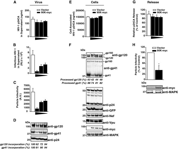
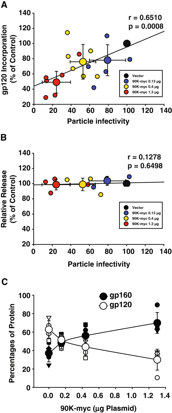
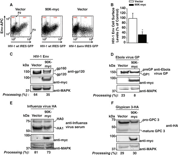
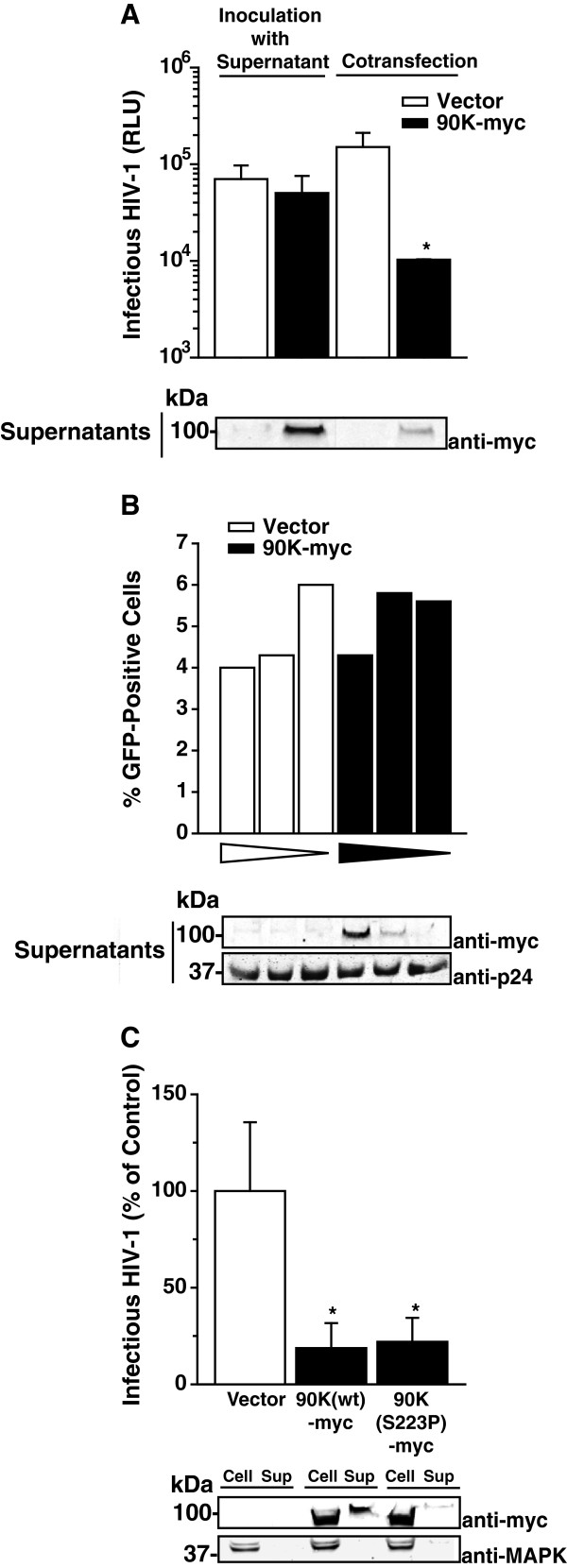
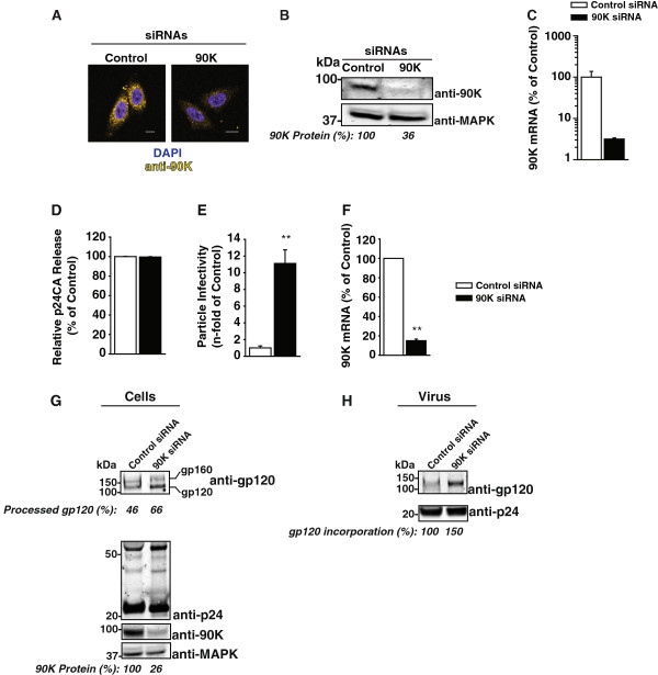
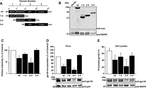
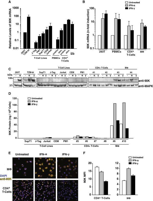
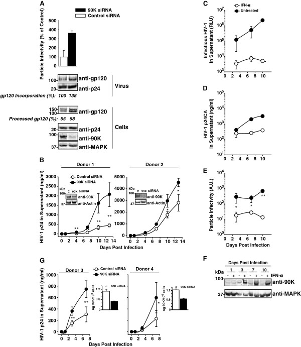
Results: Here, we show that 90K expression dose-dependently decreased the particle infectivity of HIV-1 progeny. The lower infectivity of released particles correlated with reduced virion incorporation of mature envelope glycoproteins gp120 and gp41. Further, proteolytic processing of the gp160 precursor and surface expression of gp120 in the producer cell were impaired in the presence of 90K expression. In contrast, expression of Gag, Nef and Vpu, and virus release were not grossly affected by 90K expression. 90K-imposed restriction occurred in the absence of direct interaction of 90K with HIV-1 Env or entrapment of Env in the ER. The cell-associated, but not the secreted species of 90K, mediated the antiviral effect. A truncated version of human 90K, solely consisting of the two intermediate domains, displayed a similar antiviral activity as the full-length wildtype 90K, indicating that the N-terminal SRCR-like domain and the C-terminal domain are dispensable for 90K's antiviral activity. The murine homolog of 90K, CypCAP (Cyclophilin C-associated protein), neither modulated particle infectivity of HIV-1 nor lowered the virion incorporation of mature gp120, suggesting a species-specific mode of action. 90K was expressed at basal levels in TZM-bl cells and in primary macrophages, and at low levels in CD4⁺ T-cells and PBMCs. 90K's susceptibility to IFN-mediated stimulation of expression was cell type-specific. siRNA-mediated knockdown of 90K in TZM-bl cells and primary macrophages enhanced the incorporation of Env glycoproteins into progeny virions, boosted the particle infectivity of released HIV-1, and accelerated HIV-1 spread. Conversely, treatment of HIV-1 infected macrophages with IFN-α induced 90K expression and lowered the particle infectivity of HIV-1.

Conclusions: Thus, 90K constitutes a novel antiviral factor that reduces the particle infectivity of HIV-1, involving interference with the maturation and incorporation of HIV-1 Env molecules into virions.


Figures

Figure 1
Figure 1
Heterologous expression of 90K reduces the particle infectivity of HIV-1 progeny by interfering with the maturation and incorporation of the viral glycoproteins gp120 and gp41. (A) 293T cells were cotransfected with 1.3 μg pBR.HIV-1 IRES GFP and various amounts of pcDNA6.90K-myc (1.3, 0.4, 0.15 μg) or empty vector. The total amount of DNA was kept constant (2.6 μg). Supernatants were analyzed for amounts of released HIV-1 capsid antigen by anti-p24 capsid ELISA and (B) infectious HIV-1 by applying a TZM-bl luciferase assay. (C) Particle infectivity was defined as infectivity per ng released capsid. Shown are arithmetic means ± S.D. of triplicates. (D) Sucrose cushion-purified virions were analyzed by immunoblotting. Percentages indicate the relative gp120 and gp41 incorporation, as measured by Infrared imaging-based quantification of the amount of gp120 and gp41 per p24, respectively. The values of incorporation in absence of 90K expression were set to 100%. (E) Corresponding cell lysates were analyzed for cell-associated p24 capsid by ELISA. (F) Cell lysates were analyzed by immunoblotting using the indicated antibodies. Numbers indicate the efficiency of gp120 and gp41 processing, respectively, calculated as the signal ratio of gp120 relative to (gp120 + gp160), or of gp41 relative to (gp41 + gp160), respectively. (G) Capsid release was calculated as p24 capsid antigen in the supernatant (p24Sup) to total p24 capsid (p24Cell + p24Sup). The condition without 90K was set to 100%. (H) SupT1 cells were electroporated with pBR.HIV-1 IRES GFP (10 μg) and pcDNA6.90K-myc or empty vector (10 μg). Three days post transfection, supernatants were harvested and analyzed for particle infectivity by applying a TZM-bl infectivity assay and an anti-p24 capsid ELISA of the sucrose cushion-purified supernatants. Western Blot analysis of the producer cells was performed using the indicated antibodies.
Figure 2
Figure 2
90K-induced reduction of HIV-1 particle infectivity correlates with defective gp120 incorporation into progeny, without influencing virus release. (A) Correlative analysis of gp120Env content per p24CA and HIV-1 particle infectivity (infectivity per ng p24) in the supernatant as a function of 90K plasmid amount. Small symbols represent the values of each individual transfection; large symbols represent the arithmetic means ± S.E.M. of five-six individual experiments. The Pearson’s correlation coefficient r and the corresponding p value were calculated using GraphPad Prism Software. (B) Correlative analysis of relative released virus and HIV-1 particle infectivity (infectivity per ng p24) in the supernatant as a function of 90K plasmid amount. Small symbols represent the values of each individual transfection; large symbols represent the arithmetic means ± S.E.M. of four individual experiments. The Pearson’s correlation coefficient r and the corresponding p value were calculated using GraphPad Prism Software. (C) Relative percentage of cell-associated gp120Env or gp160 Env versus total detected anti-gp120 signal as a function of cotransfected 90K plasmid amount. Values were obtained by Infrared imaging-based quantification of signals. Small symbols represent the values of each individual experiment; large symbols represent the arithmetic means ± S.E.M. of four individual experiments.
Figure 3
Figure 3
90K-myc reduces cell surface levels of HIV-1 Env and is not a global inhibitor of furin-mediated proteolytic activity. (A) 293T cells were cotransfected with pBR.HIV-1 IRES GFP and vector or pcDNA6.90K-myc. Two days post transfection, levels of surface HIV-1 gp120 were assessed by flow cytometry following staining with a human anti-HIV-1 Env antibody. Shown are representative dot plots from one experiment out of three. Red numbers indicate the MFI in gate P2 (HIV-negative cells) and P3 (HIV-positive cells). (B) Quantitative analysis of relative HIV-1 Env surface levels in the absence or presence of 90K-myc. Env cell surface levels were calculated by comparing, within the same sample, Env levels on non-GFP-expressing cells (gate P2) with Env levels on cells with medium-high expression levels (gate P3). The ratio obtained for vector-transfected cells was set to 100%. Shown is the arithmetic mean ± S.E.M. of three independent experiments. * : p < 0.05 (Student’s T-Test). (C-F) 293T cells were cotransfected with an expression vector for the indicated furin-dependent glycoproteins (1.3 μg) and vector or pcDNA6.90K-myc (1.3 μg). Two days post transfection, cells were lysed and proteins were subjected to Western Blotting using the indicated antibodies. Depicted are the efficiencies of precursor processing, measured by Infrared imaging-based quantification of the respective precursor and the cleavage product.
Figure 4
Figure 4
The cell-associated, but not the secreted species of 90K, exerts the antiviral activity. (A) 293T cells were either transfected with pBR.HIV-1 NL4.3 IRES GFP and inoculated with culture supernatant originating from vector-transfected or 90K-myc-transfected 293T cells (left half), or cotransfected with HIV-1 wt and pcDNA6.90K-myc or empty vector (right half). Two days post transfection, the amount of infectious HIV-1 in the culture supernatant was determined. Shown are the arithmetic means ± S.D. of triplicates originating from one representative experiment out of two. Supernatants of the cultures from (A) were analyzed for 90K-myc content. (B) A constant amount of HIV-1 IRES GFP virions was incubated for 15 min at 37°C with decreasing amounts of supernatant derived from 293T cells that had been transfected with empty vector or pcDNA6.90K-myc. TZM-bl were inoculated with the pretreated virus and analyzed for GFP expression three days later. Shown are the values of one representative experiment out of two. An aliquot of the pretreated inoculum that was given to the TZM-bl cells was analyzed by Western Blotting. (C) 293T cells were cotransfected with pBR.HIV-1 NL4.3 IRES GFP and indicated expression plasmids. Two days post transfection, the amount of infectious HIV-1 in the culture supernatant was determined. Values are normalized to the empty vector-cotransfected control. Shown are the arithmetic means ± S.E.M. of three independent experiments. Cell lysates and supernatants from one representive experiment were analyzed by Western Blotting. * : p < 0.05 (Student’s T-Test).
Figure 5
Figure 5
Endogenous 90K lowers the infectivity of HIV-1. (A) Confocal microscopy of TZM-bl cells immunostained for endogenous 90K and DAPI. Scale bar: 10 μm. (B) Western Blotting analysis of TZM-bl cells using an anti-90K antibody. Values indicate the relative 90K protein expression, as measured by Infrared imaging-based quantification. (C) Normalized 90K mRNA levels of the cells shown in (A). (D) Following siRNA transfection, TZM-bl cells were infected with VSV-G-pseudotyped HIV-1NL4.3 over-night, followed by thorough washing of the cells in order to remove excess virus. Three days post infection, supernatants were analyzed for released p24 capsid antigen and (E) particle infectivity, defined as infectivity per ng p24. Bars show the arithmetic means ± S.E.M. of four experiments. (F) 90K knockdown efficiencies of the three experiments shown in (D and E) were validated by quantitative RT-PCR. Bars show the arithmetic means ± S.E.M. (G) Lysates of infected cells and (H) of sucrose cushion purified viruses from one representative experiment were analyzed by Western Blotting. Percentages indicate the efficiency of gp120 processing, calculated as the signal ratio of gp120 relative to (gp120 + gp160), the relative 90K expression, calculated as the signal ratio of 90K relative to MAPK, and the relative gp120 incorporation, calculated as the amount of gp120 per p24, respectively. **: p < 0.02 (Student’s T-Test).
Figure 6
Figure 6
The two central protein-binding domains of 90K are required and sufficient for the anti-HIV-1 activity of 90K. (A) Schematic of 90K protein organization and of 90K-myc variants. (B) 293T cells were transfected with the indicated constructs, and expression was verified by Western Blotting using an anti-myc tag antibody. (C) 293T cells were cotransfected with pBR.HIV-1 NL4.3-IRES GFP and the indicated constructs. Two days post transfection, supernatants were analyzed for particle infectivity, defined as infectivity per ng p24. (D) Sucrose cushion-purified virions of three experiments were analyzed by immunoblotting. Shown is the relative gp120 incorporation, as measured by Infrared imaging-based quantification of the amount of gp120 per p24. The signal intensity in absence of 90K expression was set to 100%. (E) Cell lysates were analyzed by immunoblotting. Shown is the percentage of processed gp120, as measured by Infrared imaging-based quantification of the amount of gp120 per (gp120 + gp160). The bar diagrams show the arithmetic means ± S.E.M. of six independent experiments. **: p < 0.02 (Student’s T-Test).
Figure 7
Figure 7
Expression and susceptibility to IFN stimulation of 90K in primary HIV-1 target cells. (A) cDNA was prepared from total RNA of the indicated cell lines and primary cells, and analyzed for relative 90K mRNA expression by TaqMan-based quantitative PCR with RNaseP expression as a reference. Depicted is the relative level of 90K expression, and values obtained for 293T cells were arbitrarily set to 1. Histograms represent arithmetic means ± S.E.M. of three independent experiments, except for the data for CD4+ T-cells, which shows arithmetic means of two experiments. (B) Indicated primary cell cultures were treated with IFN-α (100 U/ml) or IFN-γ (100 U/ml) for 24-48 hours, or left untreated, prior to RNA isolation and cDNA synthesis. Shown is the fold increase of 90K mRNA level from 2-3 experiments, each performed in duplicates. (C) Western Blot analysis of indicated cells treated with IFN-α (100 U/ml) or IFN-γ (100 U/ml) for 48 hours, or left untreated. (D) Anti-90K ELISA of cell lysates, following treatment with IFN-α (100 U/ml) or IFN-γ (100 U/ml) for 48 hours where indicated. (E) Immunofluorescence stain of indicated primary cell cultures. Cells treated with IFN-α (100 U/ml) or IFN-γ (100 U/ml) for 24-48 hours where indicated or left untreated. Scale bar: 10 μm. (F) Quantification of 90K immunofluorescence by KNIME image processing plug-in and the mean fluorescence intensity (MFI) signal per cell was determined. The data represent the arithmetic mean ± S.E.M. of 154 untreated, 259 IFN-α-treated and 147 IFN-γ-treated T-cells and the mean of 85 untreated, 121 IFN-α-treated and 64 IFN-γ-treated macrophages.
Figure 8
Figure 8
Depletion of 90K in primary HIV-1 target cells enhances particle infectivity, enhances Env content of HIV-1 particles, and accelerates HIV-1 spread. (A) VSV-G/HIV-1 NL4.3-infected macrophages were transfected one day post-infection with a 90K-specific or an irrelevant siRNA. At day 7 post infection, particle infectivity of released HIV-1 was calculated as the ratio of infectivity to p24 capsid antigen. Cell lysates and sucrose cushion-purified virus were analyzed by Western Blotting. Percentages indicate the relative gp120 incorporation and the efficiency of gp120 processing, calculated as in Figure 5(G-H) (B) Macrophages obtained from two donors were siRNA-transfected and subsequently infected with HIV-1Ba-L. One day post infection, excess virus was removed by thorough washing. Supernatants were subjected to anti-p24 capsid antigen ELISA at indicated time points. Aliquots of cells were taken for 90K knockdown validation over time by Western Blotting. Shown is 90K expression at day 13 post infection. (C) Macrophages were infected with HIV-1Ba-L and simultaneously treated with IFN-α (100 U/ml). Supernatants were harvested at the indicated time points, frozen, and analyzed for released p24 capsid and (D) infectious HIV-1. Shown are arithmetic means of triplicates ± S.D. (E) Using the data from (C) and (D), the particle infectivity was calculated. Data points indicate arithmetic means of triplicates ± S.D. (F) Cell lysates were subjected to Western Blotting using indicated antibodies. (G) IL-2/PHA-stimulated PBMCs from two donors were nucleofected with a 90K-specific or an irrelevant siRNA followed by HIV-1NL4.3 infection over night. Excess virus was removed by thorough washing at indicated time points supernatants were subjected to p24 capsid antigen ELISA. 90K knockdown at the time point of infection was validated by 90K ELISA. * : p < 0.05; **: p < 0.02 (Student’s T-Test).

Similar articles

Cited by

References

    1. Sheehy AM, Gaddis NC, Choi JD, Malim MH. Isolation of a human gene that inhibits HIV-1 infection and is suppressed by the viral Vif protein. Nature. 2002;418(6898):646–650. doi: 10.1038/nature00939. - DOI - PubMed
    1. Neil SJ, Zang T, Bieniasz PD. Tetherin inhibits retrovirus release and is antagonized by HIV-1 Vpu. Nature. 2008;451(7177):425–430. doi: 10.1038/nature06553. - DOI - PubMed
    1. Van Damme N, Goff D, Katsura C, Jorgenson RL, Mitchell R, Johnson MC, Stephens EB, Guatelli J. The interferon-induced protein BST-2 restricts HIV-1 release and is downregulated from the cell surface by the viral Vpu protein. Cell Host Microbe. 2008;3(4):245–252. doi: 10.1016/j.chom.2008.03.001. - DOI - PMC - PubMed
    1. Hrecka K, Hao C, Gierszewska M, Swanson SK, Kesik-Brodacka M, Srivastava S, Florens L, Washburn MP, Skowronski J. Vpx relieves inhibition of HIV-1 infection of macrophages mediated by the SAMHD1 protein. Nature. 2011;474(7353):658–661. doi: 10.1038/nature10195. - DOI - PMC - PubMed
    1. Laguette N, Bregnard C, Benichou S, Basmaciogullari S. Human immunodeficiency virus (HIV) type-1, HIV-2 and simian immunodeficiency virus Nef proteins. Mol Aspects Med. 2010;31(5):418–433. doi: 10.1016/j.mam.2010.05.003. - DOI - PubMed

Publication types

MeSH terms