

Biological response to nano-scale titanium dioxide (TiO2): role of particle dose, shape, and retention
- PMID: 24156719
- PMCID: PMC4370163
- DOI: 10.1080/15287394.2013.826567
Biological response to nano-scale titanium dioxide (TiO2): role of particle dose, shape, and retention
Abstract
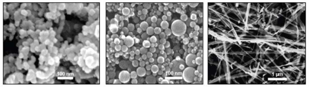
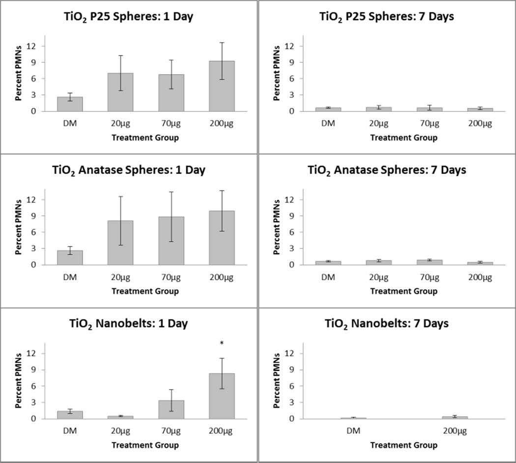
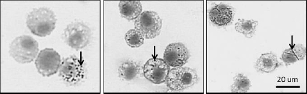
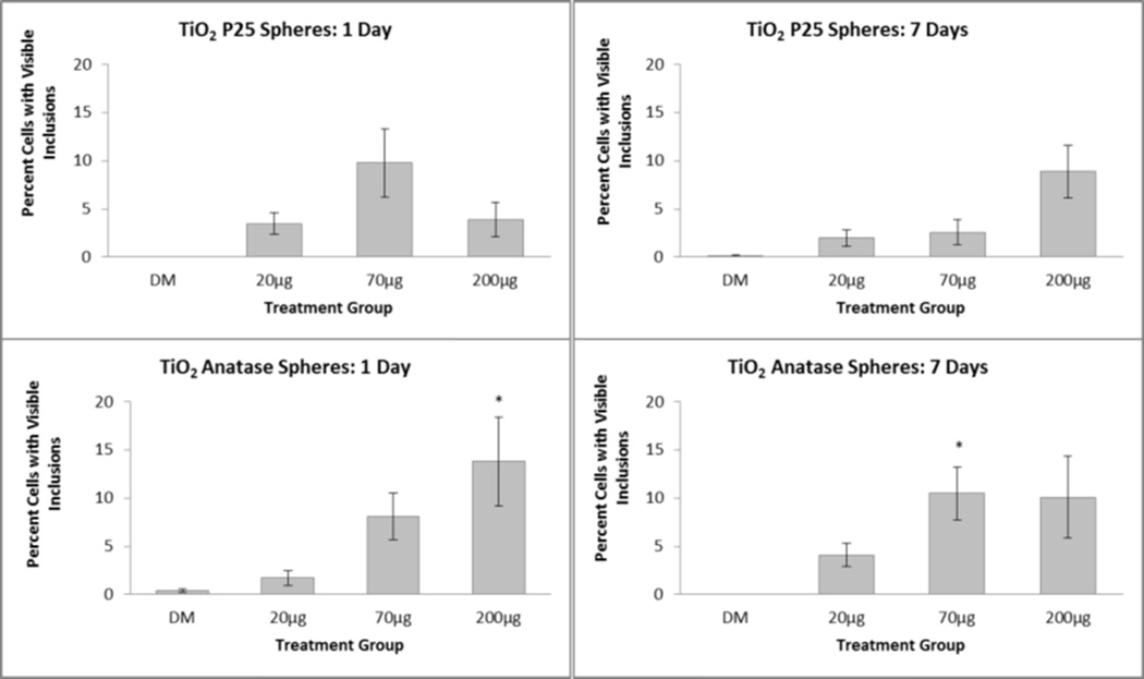
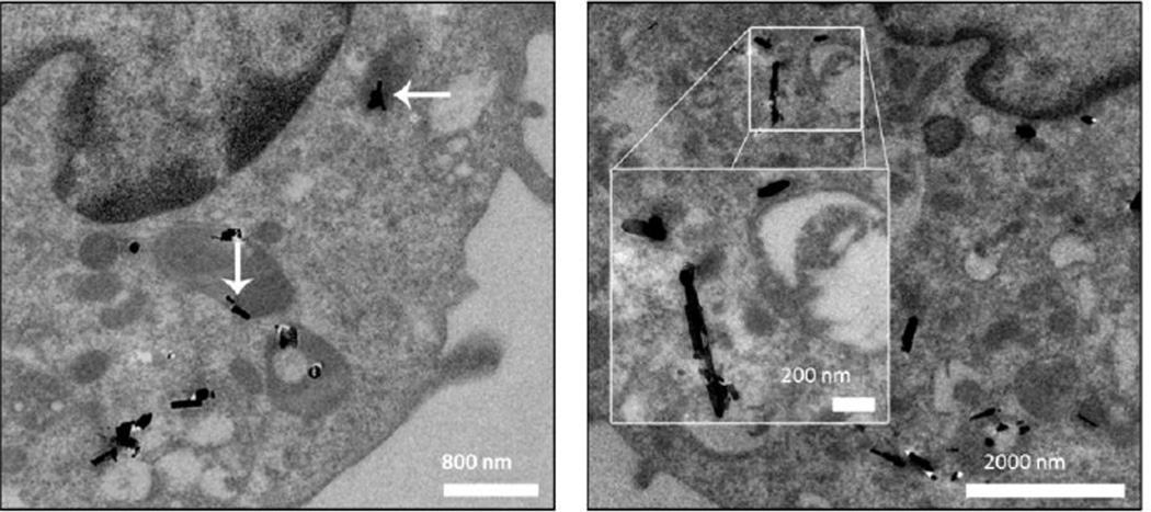
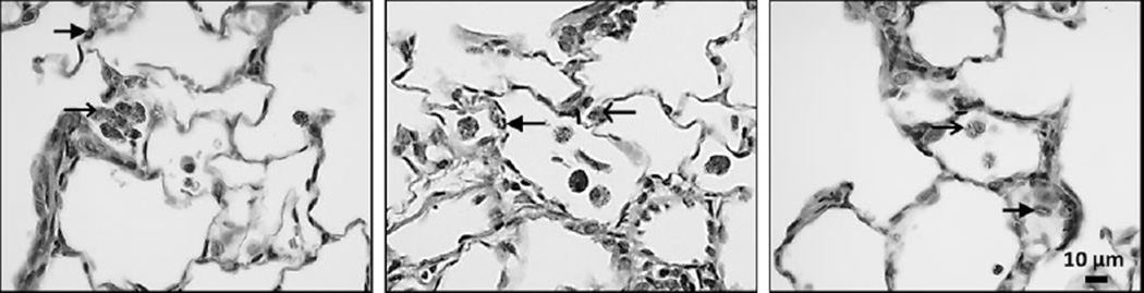
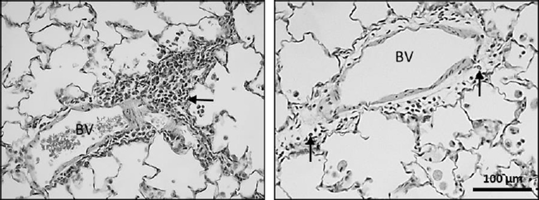
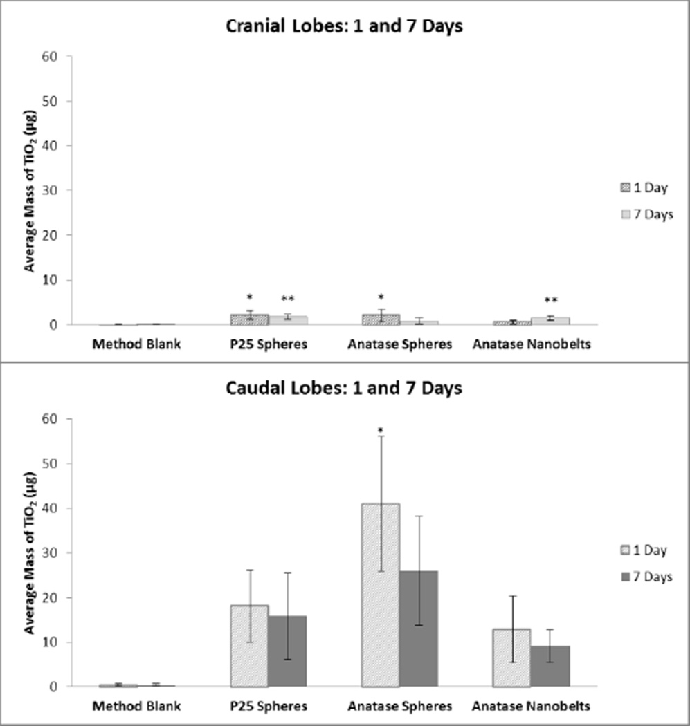
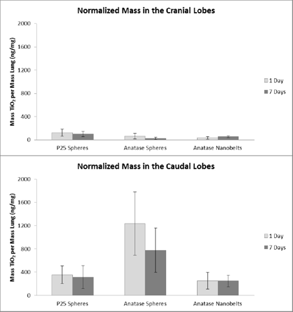
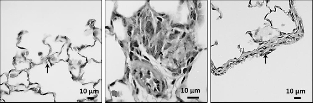
Titanium dioxide (TiO2) is one of the most widely used nanomaterials, valued for its highly refractive, photocatalytic, and pigmenting properties. TiO2 is also classified by the International Agency for Research on Cancer (IARC) as a possible human carcinogen. The objectives of this study were to (1) establish a lowest-observed-effect level (LOEL) for nano-scale TiO2, (2) determine TiO2 uptake in the lungs, and (3) estimate toxicity based on physicochemical properties and retention in the lungs. In vivo lung toxicity of nano-scale TiO2 using varying forms of well-characterized, highly dispersed TiO2 was assessed. Anatase/rutile P25 spheres (TiO2-P25), pure anatase spheres (TiO2-A), and anatase nanobelts (TiO2-NB) were tested. To determine the effects of dose and particle characteristics, male Sprague-Dawley rats were administered TiO2 (0, 20, 70, or 200 μg) via intratracheal instillation. Bronchoalveolar lavage fluid (BALF) and lung tissue were obtained for analysis 1 and 7 d post exposure. Despite abundant TiO2 inclusions in all exposed animals, only TiO2-NB displayed any significant degree of inflammation seen in BALF at the 1-d time point. This inflammation resolved by 7 d, although TiO2 particles had not cleared from alveolar macrophages recovered from the lung. Histological examination showed TiO2-NB produced cellular changes at d 1 that were still evident at d 7. Data indicate TiO2-NB is the most inflammatory with a LOEL of 200 μg at 1 d post instillation.
Figures
Similar articles
-
Interlaboratory evaluation of rodent pulmonary responses to engineered nanomaterials: the NIEHS Nano GO Consortium.Environ Health Perspect. 2013 Jun;121(6):676-82. doi: 10.1289/ehp.1205693. Epub 2013 May 6. Environ Health Perspect. 2013. PMID: 23649427 Free PMC article.
-
Pulmonary toxicity study in rats with three forms of ultrafine-TiO2 particles: differential responses related to surface properties.Toxicology. 2007 Jan 25;230(1):90-104. doi: 10.1016/j.tox.2006.11.002. Epub 2006 Nov 10. Toxicology. 2007. PMID: 17196727
-
Inhalation of titanium dioxide (P25) nanoparticles to rats and changes in surfactant protein (SP-D) levels in bronchoalveolar lavage fluid and serum.Nanotoxicology. 2019 Dec;13(10):1396-1408. doi: 10.1080/17435390.2019.1661042. Epub 2019 Sep 12. Nanotoxicology. 2019. PMID: 31512956
-
What is the impact of surface modifications and particle size on commercial titanium dioxide particle samples? - A review of in vivo pulmonary and oral toxicity studies - Revised 11-6-2018.Toxicol Lett. 2019 Mar 1;302:42-59. doi: 10.1016/j.toxlet.2018.11.008. Epub 2018 Nov 20. Toxicol Lett. 2019. PMID: 30468858 Review.
-
Risk assessment strategies for nanoscale and fine-sized titanium dioxide particles: Recognizing hazard and exposure issues.Food Chem Toxicol. 2015 Nov;85:138-47. doi: 10.1016/j.fct.2015.07.001. Epub 2015 Sep 8. Food Chem Toxicol. 2015. PMID: 26362081 Review.
Cited by
-
Acute inflammatory responses of nanoparticles in an intra-tracheal instillation rat model.PLoS One. 2015 Mar 4;10(3):e0118778. doi: 10.1371/journal.pone.0118778. eCollection 2015. PLoS One. 2015. PMID: 25738830 Free PMC article.
-
Investigating the Molecular Processes behind the Cell-Specific Toxicity Response to Titanium Dioxide Nanobelts.Int J Mol Sci. 2021 Aug 30;22(17):9432. doi: 10.3390/ijms22179432. Int J Mol Sci. 2021. PMID: 34502343 Free PMC article.
-
When 1+1>2: Nanostructured composites for hard tissue engineering applications.Mater Sci Eng C Mater Biol Appl. 2015 Dec 1;57:434-51. doi: 10.1016/j.msec.2015.07.050. Epub 2015 Aug 1. Mater Sci Eng C Mater Biol Appl. 2015. PMID: 26354283 Free PMC article. Review.
-
Aerosolized Silver Nanoparticles in the Rat Lung and Pulmonary Responses over Time.Toxicol Pathol. 2016 Jul;44(5):673-86. doi: 10.1177/0192623316629804. Epub 2016 Mar 29. Toxicol Pathol. 2016. PMID: 27025955 Free PMC article.
-
Shape-Related Toxicity of Titanium Dioxide Nanofibres.PLoS One. 2016 Mar 21;11(3):e0151365. doi: 10.1371/journal.pone.0151365. eCollection 2016. PLoS One. 2016. PMID: 26999274 Free PMC article.
References
-
- Bermudez E, Mangum JB, Wong BA, Asgharian B, Hext PM, Warheit DB, Everitt JI. Pulmonary responses of mice, rats, and hamsters to subchronic inhalation of ultrafine titanium dioxide particles. Toxicological Sciences. 2004;77(2):347–357. - PubMed
-
- Bonner JC, Silva RM, Taylor AJ, Brown JM, Hilderbrand SC, Castranova V, Porter D, Elder A, Oberdorster G, Harkema JR, Bramble LA, Kavanagh TJ, Botta D, Nel A, Pinkerton KE. Interlaboratory Evaluation of Rodent Pulmonary Responses to Engineered Nanomaterials: The NIEHS NanoGo Consortium. Environ Health Perspect. 2013 - PMC - PubMed
-
- Cui YL, Liu HT, Ze YG, Zhang ZL, Hu YY, Cheng Z, Cheng J, Hu RP, Gao GD, Wang L, Tang M, Hong FS. Gene Expression in Liver Injury Caused by Long-term Exposure to Titanium Dioxide Nanoparticles in Mice. Toxicological Sciences. 2012;128(1):171–185. - PubMed
Publication types
MeSH terms
Substances
Grants and funding
LinkOut - more resources
Full Text Sources
Other Literature Sources
Medical