. 2013 Oct 25:6:429.
doi: 10.1186/1756-0500-6-429.

Immunopurification of adenomatous polyposis coli (APC) proteins

Affiliations

Immunopurification of adenomatous polyposis coli (APC) proteins

Kerryn L Elliott et al. BMC Res Notes. .

Abstract

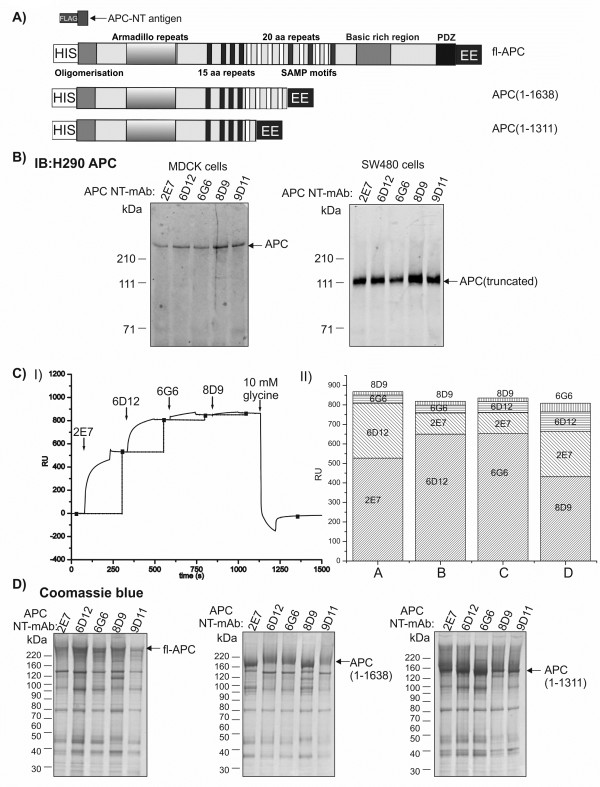
Background: The adenomatous polyposis coli (APC) tumour suppressor gene encodes a 2843 residue (310 kDa) protein. APC is a multifunctional protein involved in the regulation of β-catenin/Wnt signalling, cytoskeletal dynamics and cell adhesion. APC mutations occur in most colorectal cancers and typically result in truncation of the C-terminal half of the protein.

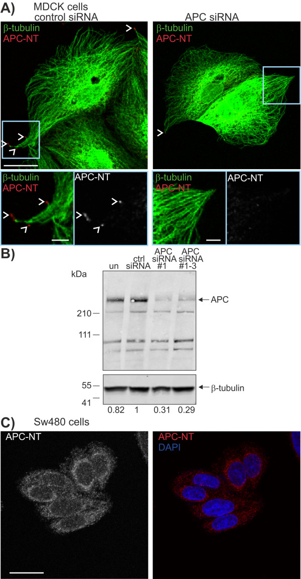
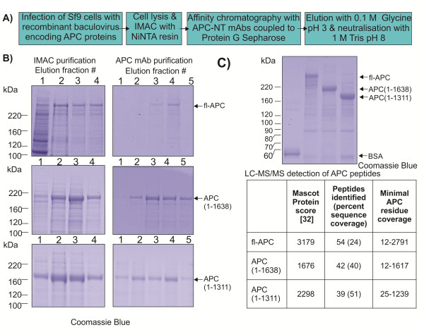
Results: In order to investigate the biophysical properties of APC, we have generated a set of monoclonal antibodies which enable purification of recombinant forms of APC. Here we describe the characterisation of these anti-APC monoclonal antibodies (APC-NT) that specifically recognise endogenous APC both in solution and in fixed cells. Full-length APC(1-2843) and cancer-associated, truncated APC proteins, APC(1-1638) and APC(1-1311) were produced in Sf9 insect cells.

Conclusions: Recombinant APC proteins were purified using a two-step affinity approach using our APC-NT antibodies. The purification of APC proteins provides the basis for detailed structure/function analyses of full-length, cancer-truncated and endogenous forms of the protein.


Figures

Figure 1
Figure 1
APC-NT mAbs recognize endogenous and recombinant full-length and truncated APC proteins in solution. A) Schematic diagram of structures of recombinant APC proteins. The APC-NT antigen (APC residues 1–61 with an N-terminal FLAG-tag) was expressed and purified from E.coli and used to generate the APC-NT mAbs. Full-length (fl-APC) and truncated recombinant APCs APC(1–1638) and APC(1–1311) were expressed in Sf9 cells with N-terminal HIS and C-terminal EE tags. The protein domains of APC are indicated: Oligomerisation, Armadillo repeats, 15 aa repeats, 20 aa repeats, SAMP motifs, basic rich domain, PDZ domain. Note: the EE-epitope tag was not used for the final affinity purification. B) Immunoprecipitation of endogenous full-length and truncated APC. APC-NT mAb immunoprecipitates from MDCK cells (1 mg protein, left panel) and SW480 colorectal carcinoma cells (1 mg protein, right panel) were immunoblotted (IB) with anti-APC H290. C) Biosensor analysis shows overlapping and non-overlapping epitopes for the APC-NT mAbs. I) Representative sensorgram showing sequential injection of APC-NT mAbs and binding to APC-NT antigen. II) Stack graph showing increases in binding (RU) to APC-NT antigen upon sequential injections with different APC-NT mAb clones. Four combinations of APC-NT mAb clones injected sequentially are represented by shaded areas (A-D). Column A corresponds to the sensorgram in I). 2E7 and 8D9 have different epitopes whereas 6D12 and 6G6 share overlapping epitopes. D) Immunoprecipitation of recombinant APC proteins expressed in Sf9 cells. APC-NT mAb (2 μg) immunoprecipitates from Sf9 cell lysates (2×10⁷ cells) expressing fl-APC (left), APC(1–1638) (middle) or APC(1–1311) (right) were resolved by SDS-PAGE and immunoprecipitated proteins were visualised with Coomassie blue. Note protein immunoprecipitated by 6D12 and 6G6 migrate slightly differently. The relative positions of the molecular markers are indicated.
Figure 2
Figure 2
APC-NT mAbs specifically detect APC at the ends of microtubules. A) MDCK cells transfected with control siRNA (top) or APC siRNA (bottom) were immunostained for APC (APC-NT mAb, 6D12 (red)) and β-tubulin (green). Distinct clusters of APC at the ends of microtubules (arrowheads) are detected by the APC-NT mAb in control but not APC depleted cells, scale bar 20 μm. Enlarged view of insets is shown (right, merged image (left) and single channel grayscale for APC (right)). Shown are single section confocal images, scale bar 5 μm. B) Depletion of cellular APC by siRNA. APC levels following siRNA transfection were assessed by immunoblot analysis (with H290 Ab from Santa Cruz) in cell lysates. Tubulin was used as a loading control. Numbers below blot are the normalised ratio of APC protein compared to control siRNA. The relative positions of molecular weight markers are indicated. C) SW480 CRC cells were immunostained for APC (APC-NT) and costained with DAPI. APC staining is diffusely cytosolic and is not detected in clusters at the ends of microtubules. Shown are single channel grayscale image for APC (left) and merged image (APC, red and DAPI blue), scale bar 20 μm.
Figure 3
Figure 3
Purification of recombinant APC proteins by a two-step method using IMAC and APC-NT mAb affinity chromatography. A) Schematic diagram depicting the purification scheme used to purify recombinant APC proteins. B) Purification of recombinant APC proteins. Sf9 cells (2×10⁹) infected with either fl-APC, APC(1–1638) or APC(1–1311) were and purified using Ni-NTA resin. Proteins were eluted using a stepwise gradient of 20, 50, 100 and 250 mM imidazole. 1% of each fraction was resolved using 4-12% Bis-Tris SDS-PAGE and stained with Coomassie blue (Left panel, 1 = 20 mM, 2 = 50 mM, 3 = 100 mM, 4 = 250 mM). Pooled fractions containing APC were applied to a 5 ml APC-NT mAb column and eluted with 0.1 M Glycine pH 3 in 5× 2 ml fractions (right). 1% of each fraction was resolved as above. The expected sizes of APC proteins are indicated by arrows and relative positions of molecular weight markers in kDa are indicated. C) Purified recombinant APC proteins. APC proteins were purified using the optimized strategy (A). The amount of APC was estimated by comparison to a band representing 1 μg BSA. The expected size of APC proteins are indicated by arrows and relative positions of molecular weight markers are indicated. Purified APC bands were excised and analysed using LC-MS/MS. MASCOT Protein score [26], peptide number, and amino acid coverage are indicated for each protein.

Similar articles

Cited by

References

    1. Fearnhead NS, Wilding JL, Bodmer WF. Genetics of colorectal cancer: hereditary aspects and overview of colorectal tumorigenesis. Br Med Bull. 2002;6:27–43. doi: 10.1093/bmb/64.1.27. - DOI - PubMed
    1. Powell SM, Zilz N, Beazer-Barclay Y, Bryan TM, Hamilton SR, Thibodeau SN, Vogelstein B, Kinzler KW. APC mutations occur early during colorectal tumorigenesis. Nature. 1992;6(6392):235–237. doi: 10.1038/359235a0. - DOI - PubMed
    1. Smith KJ, Johnson KA, Bryan TM, Hill DE, Markowitz S, Willson JK, Paraskeva C, Petersen GM, Hamilton SR, Vogelstein B. et al.The APC gene product in normal and tumor cells. Proc Natl Acad Sci U S A. 1993;6(7):2846–2850. doi: 10.1073/pnas.90.7.2846. - DOI - PMC - PubMed
    1. Hart MJ, De Los Santos R, Albert IN, Rubinfeld B, Polakis P. Downregulation of beta-catenin by human axin and its association with the APC tumor suppressor, beta-catenin and GSK3 beta. Curr Biol. 1998;6(10):573–581. doi: 10.1016/S0960-9822(98)70226-X. - DOI - PubMed
    1. Giles RH, Van Es JH, Clevers H. Caught up in a Wnt storm: Wnt signaling in cancer. Biochim Biophys Acta. 2003;6(1):1–24. - PubMed

Publication types

MeSH terms