. 2013 Oct 24;12(1):128.
doi: 10.1186/1476-4598-12-128.

A novel Hsp90 inhibitor AT13387 induces senescence in EBV-positive nasopharyngeal carcinoma cells and suppresses tumor formation

Affiliations

A novel Hsp90 inhibitor AT13387 induces senescence in EBV-positive nasopharyngeal carcinoma cells and suppresses tumor formation

King Chi Chan et al. Mol Cancer. .

Abstract

Background: Nasopharyngeal carcinoma (NPC) is an epithelial malignancy strongly associated with Epstein-Barr virus (EBV). AT13387 is a novel heat shock protein 90 (Hsp90) inhibitor, which inhibits the chaperone function of Hsp90 and reduces expression of Hsp90-dependent client oncoproteins. This study aimed to evaluate both the in vitro and in vivo antitumor effects of AT13387 in the EBV-positive NPC cell line C666-1.

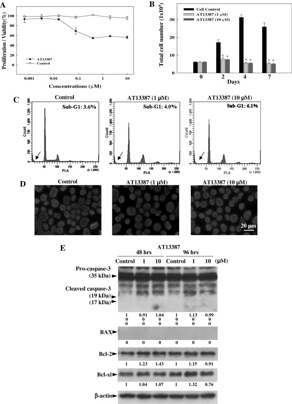
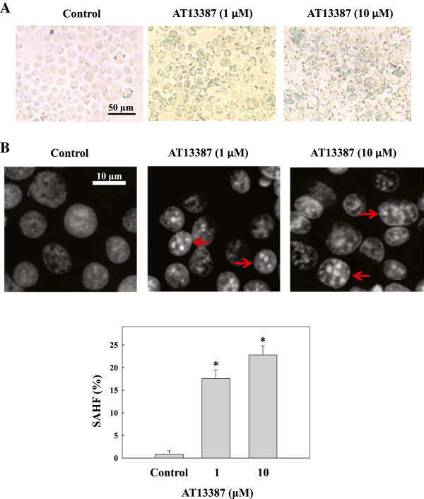
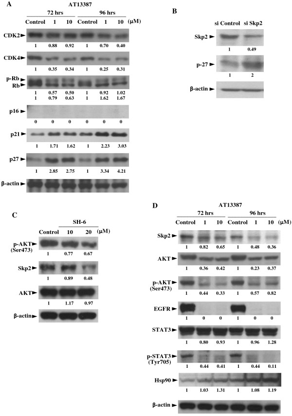
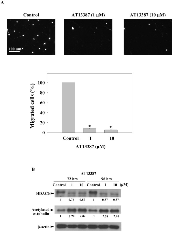
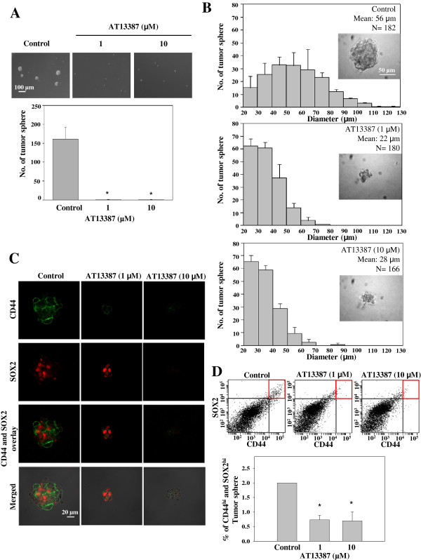
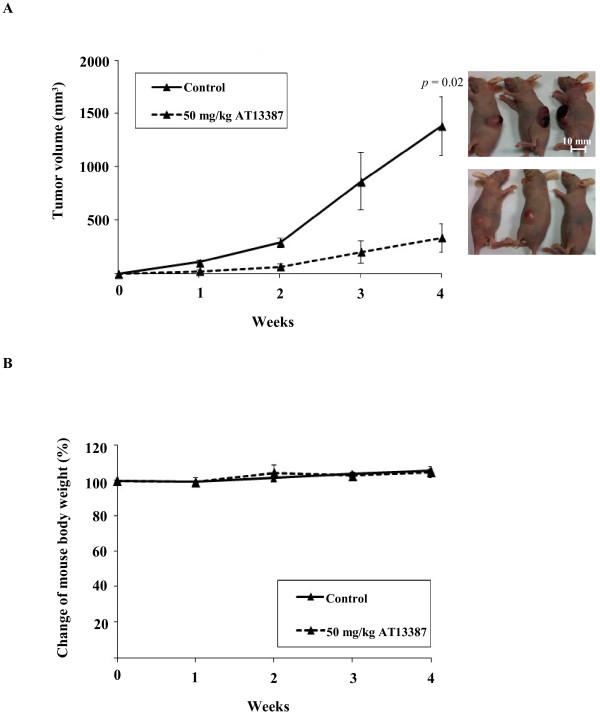
Results: Our results showed that AT13387 inhibited C666-1 cell growth and induced cellular senescence with the downregulation of multiple Hsp90 client oncoproteins EGFR, AKT, CDK4, and restored the protein expression of negative cell cycle regulator p27. We also studied the ability of AT13387 to restore p27 expression by downregulation of AKT and the p27 ubiquitin mediator, Skp2, using AKT inhibitor and Skp2 siRNA. In the functional study, AT13387 inhibited cell migration with downregulation of a cell migration regulator, HDAC6, and increased the acetylation and stabilization of α-tubulin. We also examined the effect of AT13387 on putative cancer stem cells (CSC) by 3-D tumor sphere formation assay. AT13387 effectively reduced both the number and size of C666-1 tumor spheres with decreased expression of NPC CSC-like markers CD44 and SOX2. In the in vivo study, AT13387 significantly suppressed tumor formation in C666-1 NPC xenografts.

Conclusion: AT13387 suppressed cell growth, cell migration, tumor sphere formation and induced cellular senescence on EBV-positive NPC cell line C666-1. Also, the antitumor effect of AT13387 was demonstrated in an in vivo model. This study provided experimental evidence for the preclinical value of using AT13387 as an effective antitumor agent in treatment of NPC.


Figures

Figure 1
Figure 1
Effect of AT13387 on EBV-positive NPC cell line C666-1. (A) MTT assay showing dose dependent inhibition of C666-1 cells growth after 48 hrs of AT13387 treatment. (B) Cell growth assay showing the kinetics of growth inhibition determined by counting number of viable C666-1 cells on days 2, 4 and 7 after AT13387 treatment. Results are expressed as the mean ± S.D. of three separate trials; *p < 0.05. (C) DNA content analysis and (D) DAPI nuclei staining showed no significant apoptotic phenotype in AT13387-treated C666-1 after 48 hrs. Arrow indicated the sub-G1 peak of DNA profile. Scale bar = 20 μm. (E) Western-blotting analysis showing no significant change of pro-apoptotic and anti-apoptotic proteins in AT13387-treated C666-1 after 48 hrs and 96 hrs. Value represented the arbitrary unit of band intensity after normalization with β-actin.
Figure 2
Figure 2
Characterization of senescence phenotypes in AT13387-treated C666-1. (A) Staining of senescence-associated β-galactosidase (SA-β-gal). Senescent cells were identified as cells stained blue after 72 hrs of AT13387 treatment. Scale bar = 50 μm. (B) DAPI staining and quantification of senescence-associated heterochromatic foci (SAHF). Upper panel: microscopic image showing formation of SAHF in C666-1 cells with AT13387 treatment for 96 hrs. Scale bar = 10 μm, red arrow indicated cells with typical SAHF. Lower panel: bar chart showing the percentage of cells with SAHF. At least 200 cells were counted from different microscopic fields for each treatment. Results were expressed as the mean ± S.D. of three independent experiments; *p < 0.05.
Figure 3
Figure 3
Western-blotting analysis of Hsp90 client proteins and F-box protein S-phase kinase 2 (Skp2) in AT13387-treated C666-1. (A) Expression of senescence and cell cycle associated Hsp90 client proteins in C666-1 after 72 hrs and 96 hrs AT13387 treatment. (B) Knockdown of Skp2 by siRNA leaded to upregulation of p27 in C666-1. (C) Downregulation of p-AKT (Ser473) and Skp2 by AKT inhibitor. (D) Downregulation of Skp2 and Hsp90 client oncoproteins AKT, p-AKT (Ser473), EGFR, and p-STAT3 after AT13387 treatment. Value represented the arbitrary unit of band intensity after normalization with β-actin.
Figure 4
Figure 4
Inhibitory effect of AT13387 on C666-1 cell migration. (A) Trans-well migration assay. The C666-1 cells treated with AT13387 for 72 hrs were harvested and viable cells were seeded on the upper chamber of transwell for migration assay. Upper panel: images of cells migrated through the membrane of migration chamber stained with DAPI. Scale bar = 100 μm. Lower panel: quantification of migrated cells. At least 100 cells were counted from different microscopic fields. Results were expressed as the mean ± S.D. of three separate trials; *p < 0.05. (B) Western-blotting analysis of C666-1 treated with AT13387 for 72 hrs and 96 hrs showed decreased expression of cell migration regulator HDAC6 and increased acetylated form of the α-tubulin. Value represented the arbitrary unit of band intensity after normalization with β-actin.
Figure 5
Figure 5
Effect of AT13387 on C666-1 tumor sphere. (A) AT13387 was added on day 0 of C666-1 tumor sphere formation. Upper panel: image of tumor spheres formed from C666-1 single cell culture with and without AT13387 treatment. AT13387 significantly inhibited tumor sphere formation. Scale bar = 100 μm. Lower panel: Total number of tumor spheres per culture. Tumor spheres formed with diameter reaching 20 μm were counted. Results were expressed as the mean ± S.D. of three experiments; *p < 0.05. (B) Size profile of C666-1 tumor spheres. AT13387 was added to the tumor sphere culture after tumor spheres were formed for 7 days. The diameters of tumor spheres were measured after AT13387 treatment for 7 more days and presented as the size profile of tumor sphere. The mean diameters of tumor spheres with AT13387 treatment were significantly smaller than the untreated control (p < 0.05), showing the inhibitory effect of AT13387 on the growth of C666-1 tumor sphere. Scale bar = 50 μm. Results were expressed as the mean ± S.D. of three experiments. (C) Confocal image showing pseudocolor green for CD44 and red for SOX2. Loss of CD44 was observed in 1 μM AT13387-treated C666-1 tumor sphere and loss of both CD44 and SOX2 were observed in 10 μM AT13387-treated C666-1 tumor sphere. Scale bar = 20 μm. (D) FACS analysis showing the decrease of CD44hi and SOX2hi population in AT13387-treated C666-1 tumor sphere. Results were expressed as the mean ± S.D. of three experiments; *p < 0.05.
Figure 6
Figure 6
AT13387 suppressed tumor formation in nude mouse tumorigenicity assay. (A) The tumor volume of AT13387-treated mice and vehicle control mice (n = 3) were measured weekly. The solid line represented the average tumor volume of vehicle control group, which reached 1300 mm3 in week 4. The dashed line represents the average tumor volume of AT13387-treated mice which showed significant suppression of tumor formation when compared to vehicle control. (B) The mouse body weight of AT13387-treated mice and control mice during the experiment. The result showed no significant difference for mouse body weight after AT13387 drug treatment group when compared to control group.

References

    1. Lo KW, To KF, Huang DP. Focus on nasopharyngeal carcinoma. Cancer Cell. 2004;5:423–428. doi: 10.1016/S1535-6108(04)00119-9. - DOI - PubMed
    1. Raab-Traub N. Epstein-Barr virus in the pathogenesis of NPC. Semin Cancer Biol. 2002;12:431–441. doi: 10.1016/S1044579X0200086X. - DOI - PubMed
    1. Hui AB, Cheung ST, Fong Y, Lo KW, Huang DP. Characterization of a new EBV-associated nasopharyngeal carcinoma cell line. Cancer Genet Cytogenet. 1998;101:83–88. doi: 10.1016/S0165-4608(97)00231-8. - DOI - PubMed
    1. Chan AT. Nasopharyngeal carcinoma. Ann Oncol. 2010;21(Suppl 7):vii308–vii312. - PubMed
    1. Caponigro F, Longo F, Ionna F, Perri F. Treatment approaches to nasopharyngeal carcinoma: a review. Anticancer Drugs. 2010;21:471–477. doi: 10.1097/CAD.0b013e328337160e. - DOI - PubMed

Publication types

MeSH terms