. 2013 Dec:65:137-46.
doi: 10.1016/j.yjmcc.2013.10.007. Epub 2013 Oct 21.

Tgfβ-Smad and MAPK signaling mediate scleraxis and proteoglycan expression in heart valves

Affiliations

Tgfβ-Smad and MAPK signaling mediate scleraxis and proteoglycan expression in heart valves

Damien N Barnette et al. J Mol Cell Cardiol. 2013 Dec.

Abstract

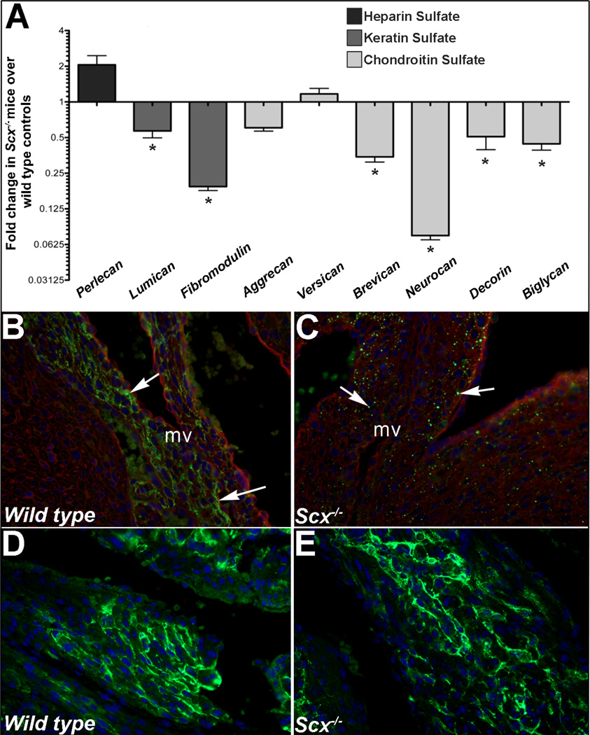
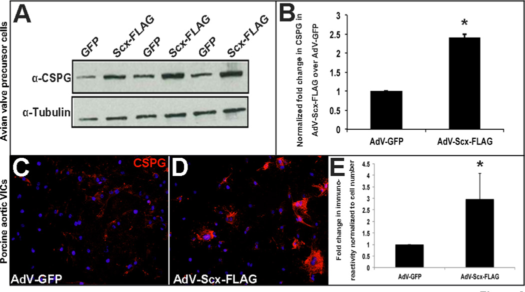
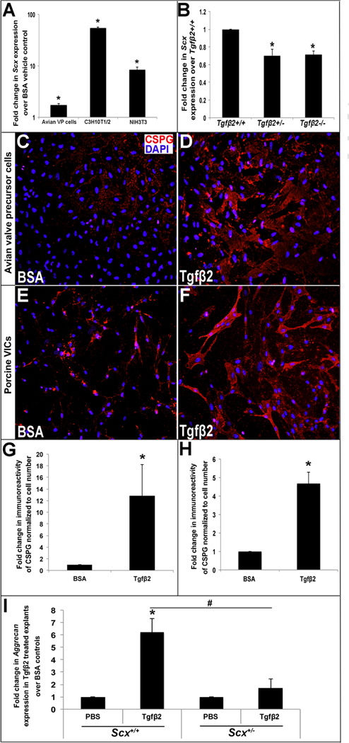
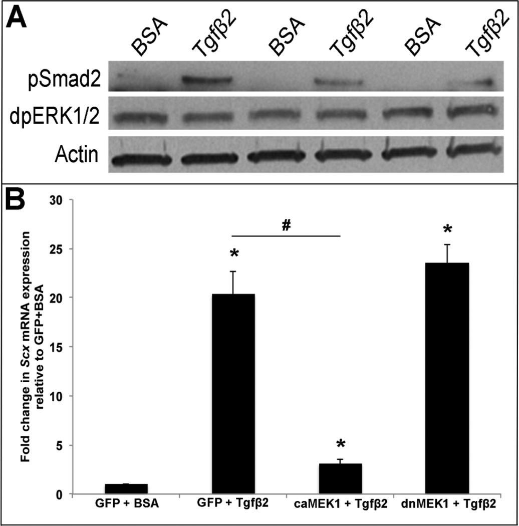
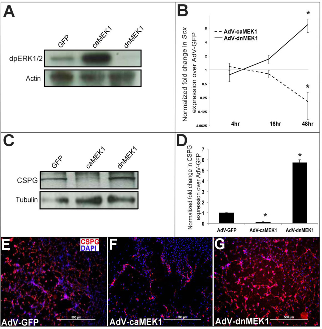
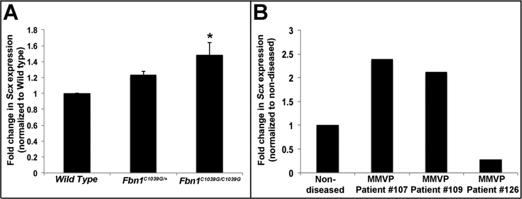
Mature heart valves are complex structures consisting of three highly organized extracellular matrix layers primarily composed of collagens, proteoglycans and elastin. Collectively, these diverse matrix components provide all the necessary biomechanical properties for valve function throughout life. In contrast to healthy valves, myxomatous valve disease is the most common cause of mitral valve prolapse in the human population and is characterized by an abnormal abundance of proteoglycans within the valve tri-laminar structure. Despite the clinical significance, the etiology of this phenotype is not known. Scleraxis (Scx) is a basic-helix-loop-helix transcription factor that we previously showed to be required for establishing heart valve structure during remodeling stages of valvulogenesis. In this study, we report that remodeling heart valves from Scx null mice express decreased levels of proteoglycans, particularly chondroitin sulfate proteoglycans (CSPGs), while overexpression in embryonic avian valve precursor cells and adult porcine valve interstitial cells increases CSPGs. Using these systems we further identify that Scx is positively regulated by canonical Tgfβ2 signaling during this process and this is attenuated by MAPK activity. Finally, we show that Scx is increased in myxomatous valves from human patients and mouse models, and overexpression in human mitral valve interstitial cells modestly increases proteoglycan expression consistent with myxomatous mitral valve phenotypes. Together, these studies identify an important role for Scx in regulating proteoglycans in embryonic and mature valve cells and suggest that imbalanced regulation could influence myxomatous pathogenesis.

Keywords: Heart valve; MAPK; Myxomatous; Proteoglycan; Scleraxis; Tgfβ.


Conflict of interest statement

Conflict of Interest. The authors of this paper have no conflicts of interest.

Figures

Figure 1
Figure 1. Proteoglycan expression is reduced in atrioventricular canal regions isolated from post natal Scx−/− mice
(A) qPCR analysis to show fold changes in proteoglycan gene expression in atrioventricular canal regions isolated from post natal Scx−/− mice compared to wild type littermate controls. * p<0.05 using Student’s t-test, n=4. (B–E) Immunohistochemistry to detect chondroitin sulfate proteoglycan (CSPG) expression (green) in mitral valves (arrows, B, C) and atria (D, E) from post natal wild type (Scx+/+)(B, D) and Scx−/−(C, E) mice. Blue indicates DAPI-stained cell nuclei, red indicates wheat germ agglutinin staining (cell membranes). mv, tricuspid valve.
Figure 2
Figure 2. Scleraxis overexpression in avian valve precursor cells and porcine valve interstitial cells promotes chondroitin sulfate proteoglycan expression
(A) Western blot analysis to show CSPG expression in HH Stage 25 avian heart valve precursor cell cultures following 48 hour infection with AdV-Scx-FLAG (Scx-FLAG) or AdV-GFP (GFP). α-Tubulin was used as a loading control. (B) Densitometry quantitation of Western blot shown in (A), *=p<0.05. (C–D) Immunohistochemistry to detect CSPG expression (red) in porcine valve interstitial (VICs) cultures infected with AdV-GFP or AdV-Scx-FLAG. Blue indicates DAPI-positive cell nuclei. (E) Quantitation of CSPG immunoreactivity shown in C–D normalized to cell number per magnification field. *=p<0.05 using Student’s t-test, n=3.
Figure 3
Figure 3. Tgfβ2 regulates Scx expression in vitro and in vivo, and promotes chondroitin sulfate proteoglycan expression
(A) qPCR analysis to show fold changes in Scx expression in avian valve precursor cells, and C3H10T1/2 and NIH3T3 murine fibroblast cell lines treated with 200pM Tgfβ2 for 48 hours compared to BSA vehicle treated controls (n=3). (B) qPCR to show Scx expression in E13.5 hearts from Tgfβ2+/− and Tgfβ2−/− mice compared to wild type (Tgfβ2+/+) littermate controls. (C–D) Immunohistochemistry to detect CSPG expression (red) in avian valve precursor cell cultures with BSA vehicle or 200pM Tgfβ2 treatment for 48 hours. (E–F) Immunohistochemistry to detect CSPG expression (red) in porcine VIC cultures treated for 48 hours with BSA vehicle or 200pM Tgfβ2. Blue indicates DAPI-positive cell nuclei. (G, H) Quantitation of CSPG immunoreactivity in avian valve precursor cells (C, D) and porcine VICs (E, F) treated with 200pM Tgfβ2 compared to BSA control. (*=p<0.05 using oneway ANOVA plus a post-hoc test n=3.) (I) qPCR to show fold changes in Aggrecan expression in mitral valve explants from Scx+/+ and Scx−/− PND1 pups treated with Tgfβ2 treatment or PBS vehicle for 48 hours. (*=p<0.05 Tgfβ2 versus PBS, #=p<0.05 Tgfβ2 treatment in Scx+/+ versus Scx−/− using Students t-test, n=3).
Figure 4
Figure 4. MEK1 activation represses Tgfβ2-mediated Scx expression
(A) Western blot analysis to show phospho-Smad2 (pSmad) and diphosho-ERK1/2 (dpERK1/2) levels in avian valve precursor cell cultures treated with 200pM Tgfβ2 for 30 minutes, compared to BSA vehicle controls. Actin was used as a loading control (B) qPCR analysis to show Scx expression in murine C3H10T1/2 cells pre-infected with AdV-GFP, AdV-caMEK1, or AdV-dnMEK1 for 6 hours prior to 48 hour treatment with 200pM Tgfβ2 or BSA vehicle control. *=p<0.05 vs. GFP+BSA, #=p<0.05 vs. GFP+Tgfβ2 using one-way ANOVA plus a post-hoc test.
Figure 5
Figure 5. Activated MEK1 signaling represses Scx and chondroitin sulfate proteoglycan expression in heart valve precursor cells
(A) Western blot analysis to show increased and decreased diphospho-ERK1/2 levels in avian heart valve precursor cells infected for 48 hours with AdV-caMEK1 and AdV-dnMEK1 respectively, compared to AdV-GFP controls. (B) qPCR to show fold changes in Scx expression in avian valve precursor cells following AdV-caMEK1 and AdV-dnMEK1 infection for 4, 16 and 48 hours, compared to AdV-GFP controls (n=4), *=p<0.05. (C) Representative Western Blot to indicate CSPG expression in avian valve precursor cells following AdV-GFP, AdV-caMEK1 and AdV-dnMEK1 treatments for 48 hours. (D) Densitometry quantitation of Western blot analysis in (C), *=p<0.05 using one-way ANOVA plus a post-hoc test. (E–G) Immunohistochemistry to detect CSPG expression in avian VP cell cultures infected with AdV-GFP (E), AdV-caMEK1 (F) or AdV-dnMEK1 (G).
Figure 6
Figure 6. Scx is increased in myxomatous mitral valves
(A) qPCR to show increased Scx expression in atrioventricular canal regions isolated from PHD6 Fbn1C1039G/C1039G mice, compared to wild type littermate controls. (B) qPCR to show changes in Scx expression in mitral VICs isolated from human patients diagnosed with myxomatous mitral valve disease, compared to mitral VICs collected from four, non-diseased hearts. *=p<0.05 using one-way ANOVA plus a post-hoc test.

References

    1. Lincoln J, Yutzey KE. Molecular and developmental mechanisms of congenital heart valve disease. Birth defects research Part A, Clinical and molecular teratology. 2011;91:526–534. - PubMed
    1. Guy TS, Hill AC. Mitral valve prolapse. Annual review of medicine. 2012;63:277–292. - PubMed
    1. Nasuti JF, Zhang PJ, Feldman MD, Pasha T, Khurana JS, Gorman JH, 3rd, et al. Fibrillin and other matrix proteins in mitral valve prolapse syndrome. The Annals of thoracic surgery. 2004;77:532–536. - PubMed
    1. Akhtar S, Meek KM, James V. Ultrastructure abnormalities in proteoglycans, collagen fibrils, and elastic fibers in normal and myxomatous mitral valve chordae tendineae. Cardiovascular pathology : the official journal of the Society for Cardiovascular Pathology. 1999;8:191–201. - PubMed
    1. Olsen EG, Al-Rufaie HK. The floppy mitral valve. Study on pathogenesis. British heart journal. 1980;44:674–683. - PMC - PubMed

Publication types

Substances