. 2014;6(2):219-26.
doi: 10.1159/000354984. Epub 2013 Oct 18.

Mast cell-deficient kit mice develop house dust mite-induced lung inflammation despite impaired eosinophil recruitment

Affiliations

Mast cell-deficient kit mice develop house dust mite-induced lung inflammation despite impaired eosinophil recruitment

J Daan de Boer et al. J Innate Immun. 2014.

Abstract

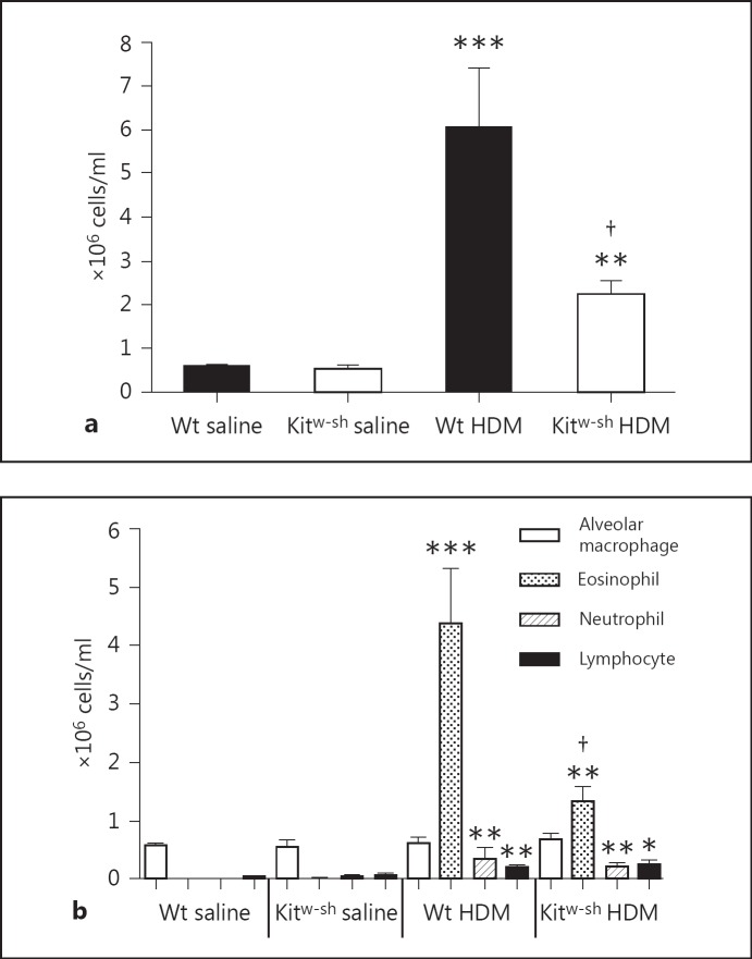
Background: Mast cells are implicated in allergic and innate immune responses in asthma, although their role in models using an allergen relevant for human disease is incompletely understood. House dust mite (HDM) allergy is common in asthma patients. Our aim was to investigate the role of mast cells in HDM-induced allergic lung inflammation.

Methods: Wild-type (Wt) and mast cell-deficient Kit(w-sh) mice on a C57BL/6 background were repetitively exposed to HDM via the airways.

Results: HDM challenge resulted in a rise in tryptase activity in bronchoalveolar lavage fluid (BALF) of Wt mice, indicative of mast cell activation. Kit(w-sh) mice showed a strongly attenuated HDM- induced recruitment of eosinophils in BALF and lung tissue, accompanied by reduced pulmonary levels of the eosinophil chemoattractant eotaxin. Remarkably, Kit(w-sh) mice demonstrated an unaltered capacity to develop lung pathology and increased mucus production in response to HDM. The increased plasma IgE in response to HDM in Wt mice was absent in Kit(w-sh) mice.

Conclusion: These data contrast with previous reports on the role of mast cells in models using ovalbumin as allergen in that C57BL/6 Kit(w-sh) mice display a selective impairment of eosinophil recruitment without differences in other features of allergic inflammation.


Figures

Fig. 1
Fig. 1
Tryptase activity levels (ΔOD vs. OD at baseline): HDM airway exposure induces tryptase activity in BALF of Wt but not of Kitw-sh mice. Data are means ± SEM of 8 mice per group. ** p < 0.01 versus saline; p < 0.05 versus HDM-exposed Wt mice. OD = Optical density.
Fig. 2
Fig. 2
Kitw-sh mice have decreased total cell counts in BALF after HDM challenge due to lower eosinophil influx: total cell counts (a) and differential cell counts (alveolar macrophages, eosinophils, neutrophils and lymphocytes; b). Data are means ± SEM (106 cells/ml) of 8 mice per group. * p < 0.05, ** p < 0.01 and *** p < 0.001 versus saline-challenged mice of the same genotype; p < 0.05 versus Wt mice challenged with HDM.
Fig. 3
Fig. 3
Kitw-sh mice demonstrate a reduced influx of eosinophils in lung tissue after HDM challenge. a Percentage of lung surface stained positive for eosinophils quantified by digital imaging of GMBP staining (see Materials and Methods). Data are means ± SEM of 8 mice per group except for Wt saline (n = 5). * p < 0.05 and ** p < 0.01 versus saline-challenged mice of the same genotype; p < 0.05 versus Wt mice challenged with HDM. b Representative GMBP staining of lung tissue slides of Wt mice exposed to saline, Kitw-sh mice exposed to saline, Wt mice exposed to HDM and Kitw-sh mice exposed to HDM. Original magnification ×40.
Fig. 4
Fig. 4
Kitw-sh and Wt mice demonstrate similar lung pathology after HDM challenge. a Semiquantitative pathology scores (described in Materials and Methods). Data are means ± SEM of 8 mice per group except for Wt saline (n = 5). ** p < 0.01 and *** p < 0.001 versus saline-challenged mice of the same genotype. b Representative HE-stained lung tissue slides of Wt mice exposed to saline, Kitw-sh mice exposed to saline, Wt mice exposed to HDM and Kitw-sh mice exposed to HDM. Original magnification ×40.
Fig. 5
Fig. 5
Kitw-sh mice show unaltered mucus production in their airways upon HDM challenge. a Semiquantitative mucus scores (described in Materials and Methods). Data are means ± SEM of 8 mice per group except for Wt saline (n = 5). ** p < 0.01 and *** p < 0.001 versus saline-challenged mice of the same genotype. b Representative PAS-D-stained lung tissue slides of Wt mice exposed to saline, Kitw-sh mice exposed to saline, Wt mice exposed to HDM and Kitw-sh mice exposed to HDM. Original magnification ×40.
Fig. 6
Fig. 6
Lung concentrations of eotaxin: pulmonary eotaxin levels are decreased in Kitw-sh mice after HDM exposure. Data are means ± SEM of 8 mice per group except for Wt saline (n = 5). * p < 0.05 versus saline-challenged Wt mice; p < 0.05 versus HDM-challenged Wt mice.
Fig. 7
Fig. 7
Plasma concentrations of IgE: Kitw-sh mice do not show a plasma IgE response after HDM challenge. Data are means ± SEM of 8 mice per group. * p < 0.05 versus saline-challenged mice.

Similar articles

Cited by

References

    1. Braman SS. The global burden of asthma. Chest. 2006;130:4S–12S. - PubMed
    1. Murphy DM, O'Byrne PM. Recent advances in the pathophysiology of asthma. Chest. 2010;137:1417–1426. - PubMed
    1. Gregory LG, Lloyd CM. Orchestrating house dust mite-associated allergy in the lung. Trends Immunol. 2011;32:402–411. - PMC - PubMed
    1. Nelson RP, Jr, DiNicolo R, Fernandez-Caldas E, Seleznick MJ, Lockey RF, Good RA. Allergen-specific IgE levels and mite allergen exposure in children with acute asthma first seen in an emergency department and in nonasthmatic control subjects. J Allergy Clin Immunol. 1996;98:258–263. - PubMed
    1. Lodge CJ, Lowe AJ, Gurrin LC, Hill DJ, Hosking CS, Khalafzai RU, Hopper JL, Matheson MC, Abramson MJ, Allen KJ, Dharmage SC. House dust mite sensitization in toddlers predicts current wheeze at age 12 years. J Allergy Clin Immunol. 2011;128:782–788. e9. - PubMed

Publication types

MeSH terms