. 2013 Nov 8;342(6159):731-4.
doi: 10.1126/science.1242059. Epub 2013 Oct 24.

High-resolution mapping of the spatial organization of a bacterial chromosome

Affiliations

High-resolution mapping of the spatial organization of a bacterial chromosome

Tung B K Le et al. Science. .

Abstract

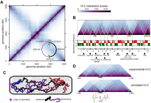
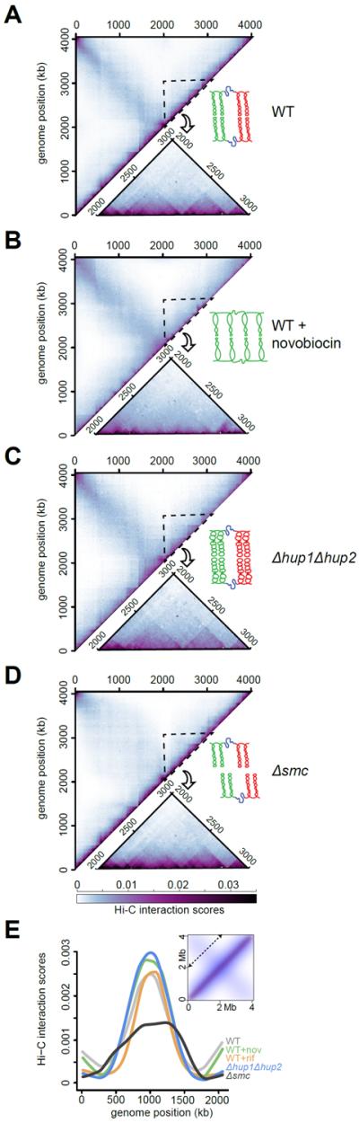
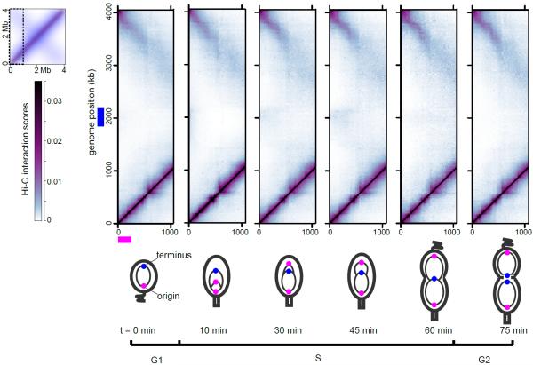
Chromosomes must be highly compacted and organized within cells, but how this is achieved in vivo remains poorly understood. We report the use of chromosome conformation capture coupled with deep sequencing (Hi-C) to map the structure of bacterial chromosomes. Analysis of Hi-C data and polymer modeling indicates that the Caulobacter crescentus chromosome consists of multiple, largely independent spatial domains that are probably composed of supercoiled plectonemes arrayed into a bottle brush-like fiber. These domains are stable throughout the cell cycle and are reestablished concomitantly with DNA replication. We provide evidence that domain boundaries are established by highly expressed genes and the formation of plectoneme-free regions, whereas the histone-like protein HU and SMC (structural maintenance of chromosomes) promote short-range compaction and the colinearity of chromosomal arms, respectively. Collectively, our results reveal general principles for the organization and structure of chromosomes in vivo.


Figures

Fig. 1
Fig. 1. Partitioning of the Caulobacter chromosome into chromosomal interaction domains (CIDs)
(A) Normalized NcoI Hi-C contact map for Caulobacter swarmer cells displaying contact frequencies for pairs of 10-kb bins across the genome. Axes indicate the genome position of each bin. Inset: simplified genomic map showing the origin of replication (ori) and terminus (ter) along with the right (black) and left (grey) chromosomal arms. (B) Hi-C contact map for one arm of the chromosome rotated 45° clockwise with directional preference plots below. Left- and right-ward preferences are shown as green and red bars, respectively. CIDs are outlined in yellow and numbered. Highly-expressed genes at CID boundaries are listed (hypothetical genes designated by GenBank ID). (C) Polymer chromosome model indicating polarly-anchored origin (magenta), chromosome backbone (black), and plectonemes (grey, with every 10th plectoneme on one arm in a color). (D) Comparison of experimental and simulated Hi-C contact maps, indicating that plectoneme-free regions (PFRs) can account for CIDs.
Fig. 2
Fig. 2. Effect of inhibiting transcription on CID boundaries
Normalized BglII Hi-C contact maps for (A) untreated and (B) rifampicin-treated swarmer cells. (C) Hi-C contact maps for wild-type, ΔrsaA, and ΔrsaA + van::PrsaA-rsaA. Only the region of the genome containing the van locus (dashed line) is shown.
Fig. 3
Fig. 3. Contribution of supercoiling, HU, and SMC to chromosome organization
Normalized BglII Hi-C contact maps for (A) wild type, (B) novobiocin-treated wild type, (C) Δhup1Δhup2, and (D) Δsmc swarmer cells. Only the upper left regions of the symmetric Hi-C maps are shown. A region from 2–3 Mb is enlarged and shown below each map. A cartoon summarizing the effects of each perturbation is shown with neighboring domains of plectonemic DNA in red and green and plectoneme-free regions in blue. (E) Hi-C scores for the diagonal (indicated in inset) of each contact map.
Fig. 4
Fig. 4. Dynamics of chromosomal organization during cell cycle progression
Plots show a section of the Hi-C interaction map for the cell-cycle time points indicated.

References

    1. Booker BM, Deng S, Higgins NP. Mol Microbiol. 2010;78:1348. - PubMed
    1. Fisher JK, et al. Cell. 2013;153:882. - PMC - PubMed
    1. Niki H, Yamaichi Y, Hiraga S. Genes Dev. 2000;14:212. - PMC - PubMed
    1. Umbarger MA, et al. Mol Cell. 2011;44:252. - PMC - PubMed
    1. Valens M, Penaud S, Rossignol M, Cornet F, Boccard F. EMBO J. 2004;23:4330. - PMC - PubMed

Publication types

MeSH terms

Associated data