Comparative Study
. 2013 Nov;22(13):1371-81.
doi: 10.1177/0961203313508444.

Differential immunoglobulin class-mediated responses to components of the U1 small nuclear ribonucleoprotein particle in systemic lupus erythematosus and mixed connective tissue disease

Affiliations
Comparative Study

Differential immunoglobulin class-mediated responses to components of the U1 small nuclear ribonucleoprotein particle in systemic lupus erythematosus and mixed connective tissue disease

A Mesa et al. Lupus. 2013 Nov.

Abstract

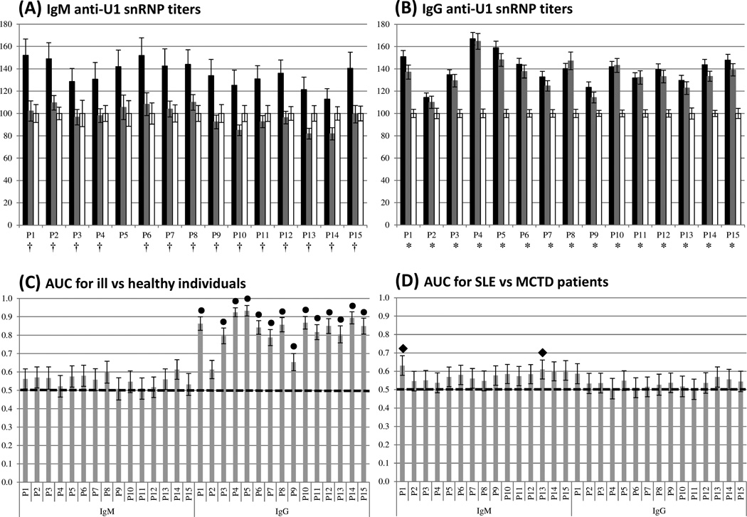
Objective: The objective of this paper is to determine whether patients with systemic lupus erythematosus (SLE) and mixed connective tissue disease (MCTD) possess differential IgM- and IgG-specific reactivity against peptides from the U1 small nuclear ribonucleoprotein particle (U1 snRNP).

Methods: The IgM- and IgG-mediated responses against 15 peptides from subunits of the U1 snRNP were assessed by indirect enzyme linked immunosorbent assays (ELISAs) in sera from patients with SLE and MCTD and healthy individuals (n = 81, 41, and 31, respectively). Additionally, 42 laboratory tests and 40 clinical symptoms were evaluated to uncover potential differences. Binomial logistic regression analyses (BLR) were performed to construct models to support the independent nature of SLE and MCTD. Receiver operating characteristic (ROC) curves corroborated the classification power of the models.

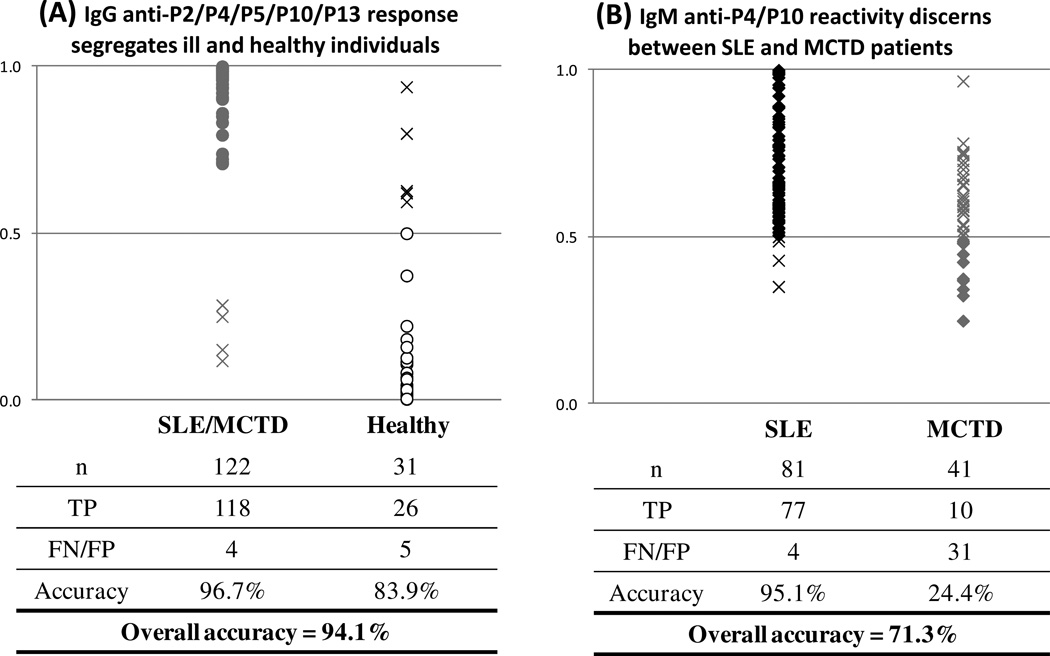
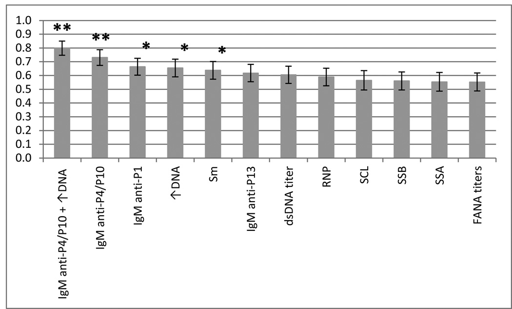
Results: We analyzed IgM and IgG anti-U1 snRNP titers to classify SLE and MCTD patients. IgG anti-U1 snRNP reactivity segregates SLE and MCTD from nondisease controls with an accuracy of 94.1% while IgM-specific anti-U1 snRNP responses distinguish SLE from MCTD patients with an accuracy of 71.3%. Comparison of the IgG and IgM anti-U1 snRNP approach with clinical tests used for diagnosing SLE and MCTD revealed that our method is the best classification tool of those analyzed (p ≤ 0.0001).

Conclusions: Our IgM anti-U1 snRNP system along with lab tests and symptoms provide additional molecular and clinical evidence to support the hypothesis that SLE and MCTD may be distinct syndromes.

Keywords: Systemic lupus erythematosus (SLE); U1 small nuclear ribonucleoprotein particle (U1 snRNP); autoimmune disorders; classification criteria; immunoglobulin M (IgM); mixed connective tissue disease (MCTD).


Figures

Figure 1
Figure 1. Contrasting IgM-specific anti-U1 snRNP peptide responses observed in SLE and MCTD patients
(A) and (B) represent the average percent optical density (OD%) values for the IgM class and IgG classes, respectively. Peptide number and OD% are on the x and y axes, respectively. The black, gray and white bars symbolize the average OD% of SLE, MCTD and healthy groups, respectively. (†) and (*) indicate significantly different OD% between SLE and MCTD as well as patients and healthy populations, respectively (p ≤ 0.05). (C) and (D) correspond to the area under the curve (AUC), derived from ROC curves, for ill (SLE and MCTD) vs. healthy individuals as well as SLE vs. MCTD patients, respectively. Peptide number per Ig class and their AUC values are indicated on the x and y axes, respectively. (●) and (♦) symbolize significantly different AUC between patients and healthy individuals as well as SLE and MCTD patients, respectively (p ≤ 0.05). The dotted lines in C and D indicate the cut-off value (0.5). Black bars in all graphs represent standard error of the mean.
Figure 2
Figure 2. Identification of a two-step ELISA system for classification of SLE, MCTD and healthy individuals
(A) The combination of IgG-mediated anti-P2/P4/P5/P10/P13 provides the best segregation between SLE and MCTD and non-disease controls. The distribution of Ill (SLE and MCTD) and healthy individuals and the predicted combined IgG-mediated reactivity are represented on the x and y axes, respectively. Gray and white circles indicate true positives (TP). (B) Combined IgM-anti-P4/P10 can classify SLE and MCTD patients. The distribution of SLE and MCTD patients’ combined IgM anti-P4/P10 predicted values are on the x and y axes, respectively. Black and gray diamonds indicate true positive (TP) samples for SLE and MCTD patients, respectively. The crosses represent false negatives (FN) or false positives (FP). Predicted values were obtained using binomial logistic regression (BLR) with a cut-off of 0.5 (p ≤ 0.05).
Figure 3
Figure 3. Area under the curve analysis reveals the classification power of IgM anti-P4/P10
Receiver operating characteristic (ROC) curves were generated using peptide antigenicities or laboratory tests. The columns in the graph represent the area under the curve (AUC) on the y axis for each variable tested. The bars on top of each column indicate standard error of the mean. FANA titers, dsDNA, ↑DNA (elevated serum DNA) and positive results for RNP, Sm, SSA, SSB and SCL are clinical tests used during SLE and MCTD diagnosis. The “IgM anti-P4/P10” indicates the combined IgM anti-P4/P10 titer while “IgM anti-P4/P10 + ↑DNA” represents the combination of the IgM anti-P4/P10 ELISA and the elevated DNA assay. The “*” and “**” indicate significant differences in classifying SLE and MCTD with p values of ≤ 0.05 and ≤ 0.0001, respectively.

References

    1. Riemekasten G, Hahn BH. Key autoantigens in SLE. Rheumatol (Oxford) 2005;44:975–982. - PubMed
    1. Liu CC, Ahearn JM. The search for lupus biomarkers. Best Pract Res Clin Rheumatol. 2009;4:507–523. - PMC - PubMed
    1. Zdrojewicz Z, Budzyń-Kozioł E, Puławska J. Mixed connective tissue disease--etiology, pathogenesis, clinical significance, treatment. Postepy Hig Med Dosw. 1999;53:751–766. - PubMed
    1. Egner W. The use of laboratory tests in the diagnosis of SLE. J Clinical Pathol. 2000;53:424–432. - PMC - PubMed
    1. Greidinger EL, Hoffman RW. The appearance of U1 RNP antibody specificities in sequential autoimmune human antisera follows a characteristic order that implicates the U1-70 kd and B'/B proteins as predominant U1 RNP immunogens. Arthritis Rheum. 2001;44:368–375. - PubMed

Publication types

MeSH terms