. 2013 Oct 25;342(6157):1241006.
doi: 10.1126/science.1241006.

Fine tuning of craniofacial morphology by distant-acting enhancers

Affiliations

Fine tuning of craniofacial morphology by distant-acting enhancers

Catia Attanasio et al. Science. .

Abstract

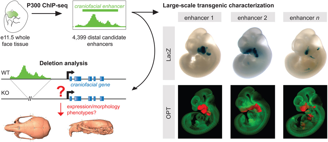
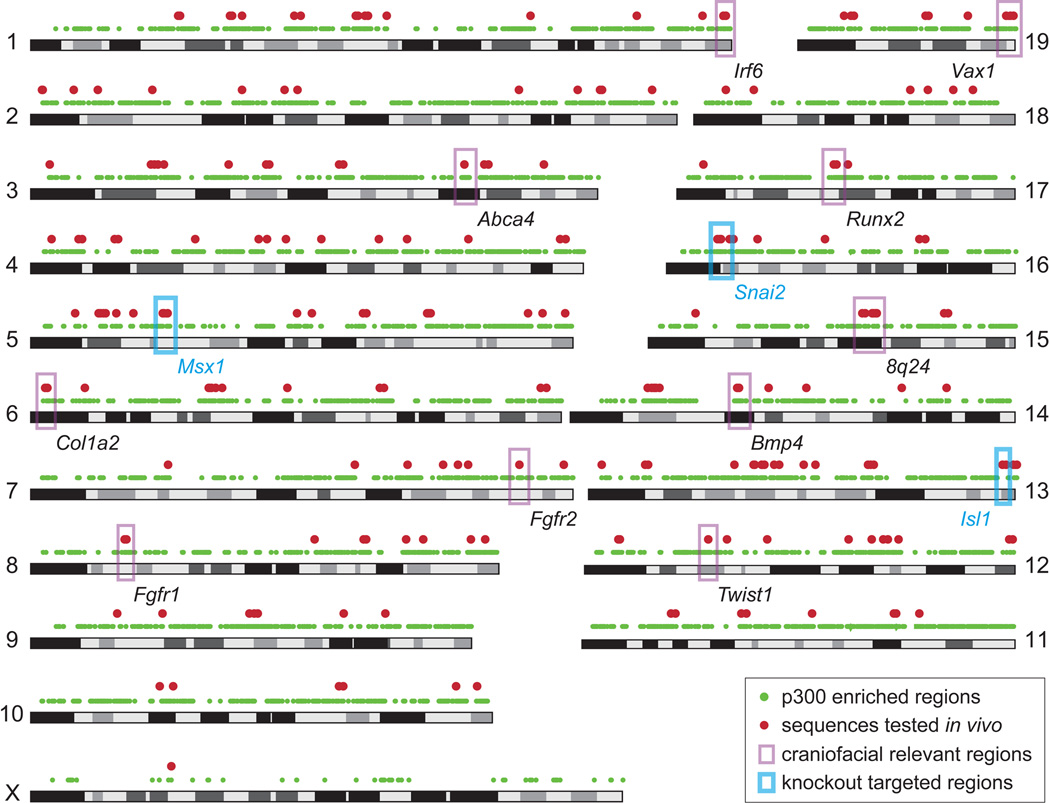
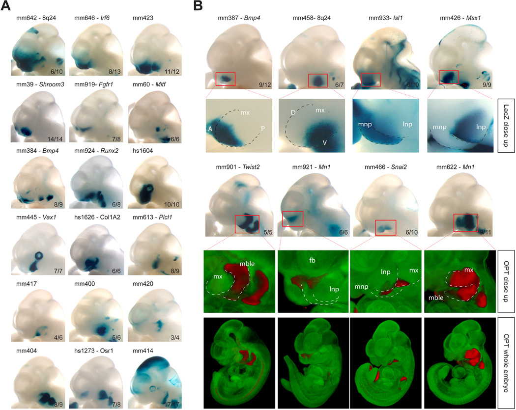
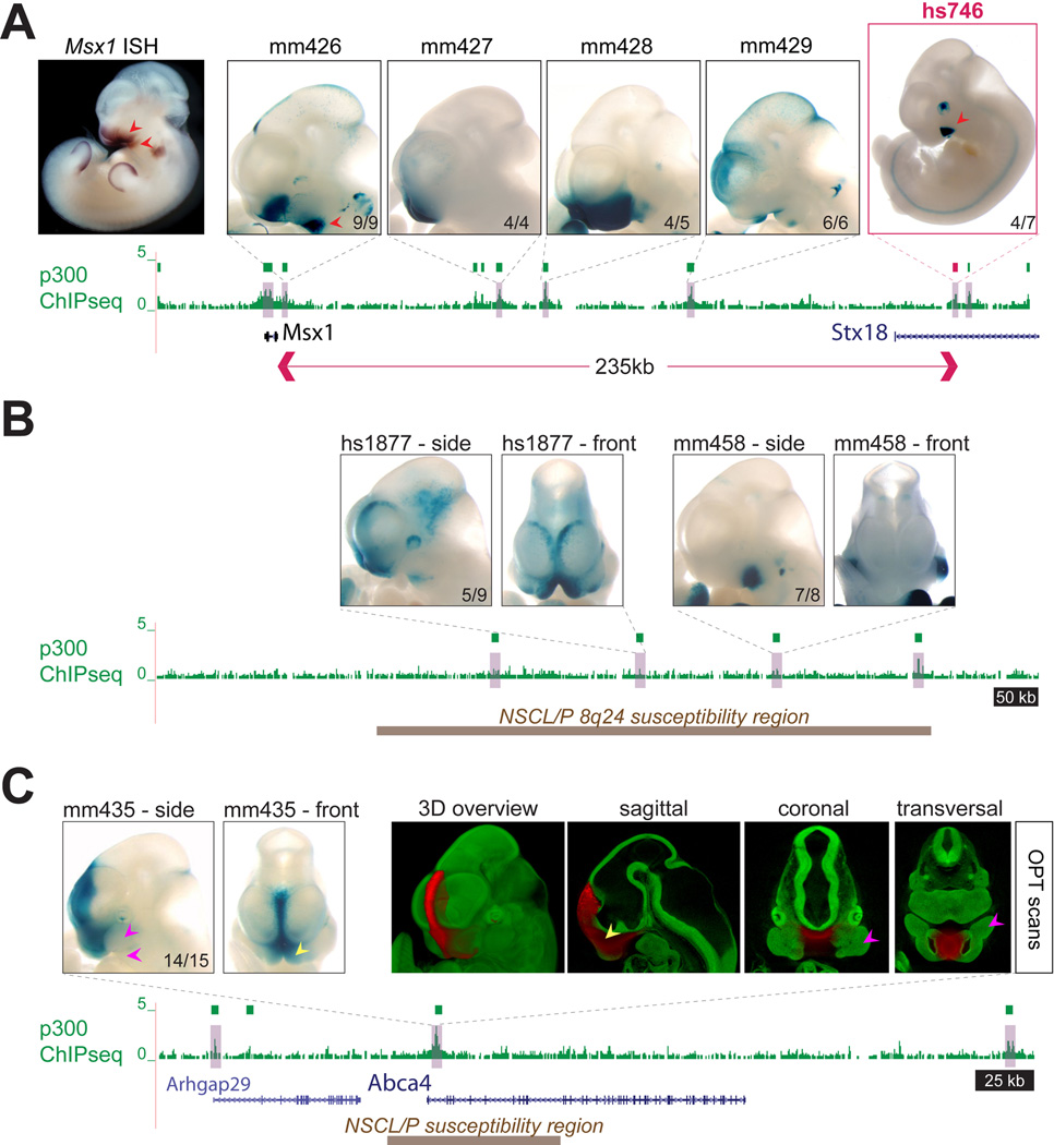
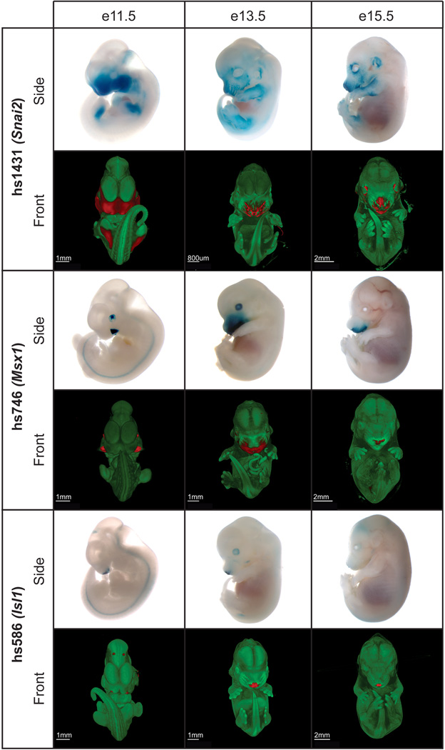
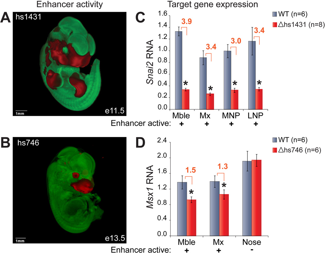
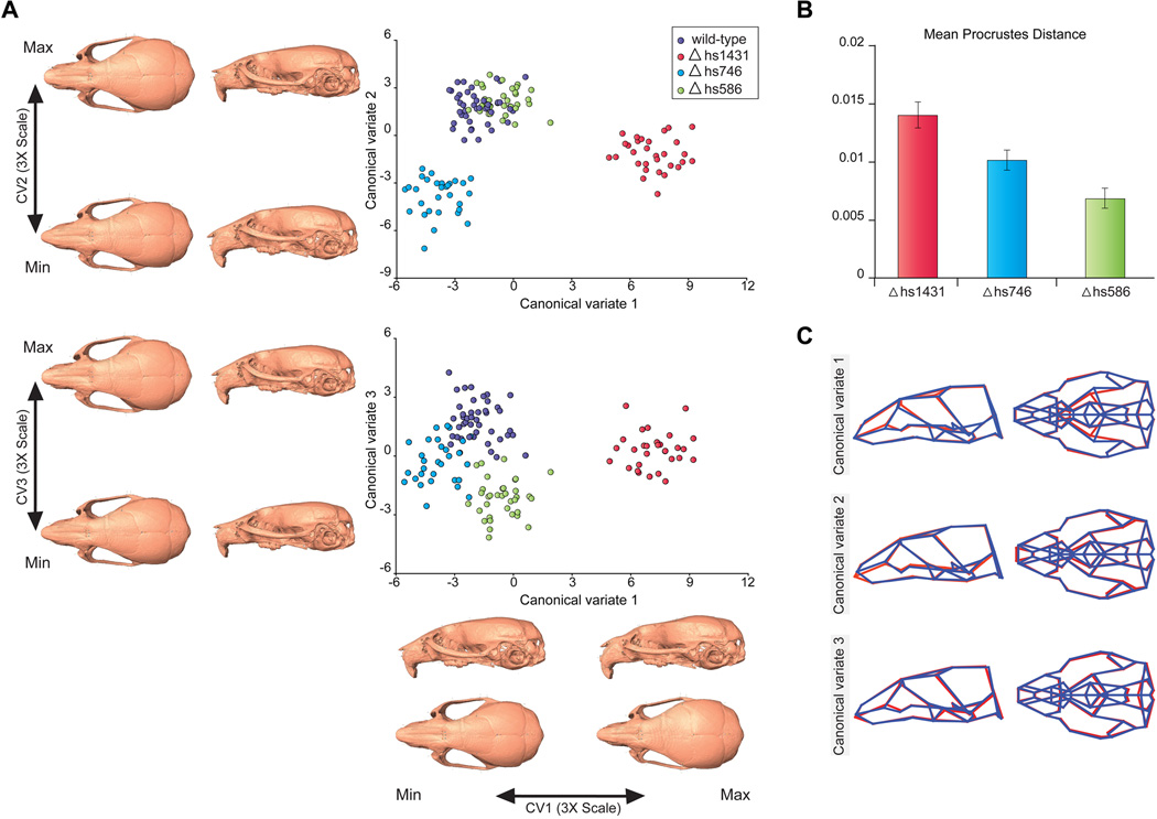
The shape of the human face and skull is largely genetically determined. However, the genomic basis of craniofacial morphology is incompletely understood and hypothesized to involve protein-coding genes, as well as gene regulatory sequences. We used a combination of epigenomic profiling, in vivo characterization of candidate enhancer sequences in transgenic mice, and targeted deletion experiments to examine the role of distant-acting enhancers in craniofacial development. We identified complex regulatory landscapes consisting of enhancers that drive spatially complex developmental expression patterns. Analysis of mouse lines in which individual craniofacial enhancers had been deleted revealed significant alterations of craniofacial shape, demonstrating the functional importance of enhancers in defining face and skull morphology. These results demonstrate that enhancers are involved in craniofacial development and suggest that enhancer sequence variation contributes to the diversity of human facial morphology.


Figures

Fig. 1
Fig. 1. Study Overview
P300 ChIP-seq was performed on whole mouse face tissue from e11.5 embryos, which identified 4,399 putative distant-acting craniofacial enhancers. More than 200 craniofacial candidate enhancers were characterized in depth through LacZ transgenesis in mouse embryos (LacZ panel), and selected enhancers were further analyzed by optical projection tomography (OPT panel; unstained tissue is shown in green, LacZ stained tissue is shown in red). Furthermore, a panel of three enhancers near functionally unrelated genes was studied by knockout analysis and detailed skull morphometry in mice.
Fig. 2
Fig. 2. Genome-wide identification of candidate craniofacial enhancers
Mouse genome graph showing all p300-enriched regions (green dots) and all 281 sequences tested in vivo or re-examined for craniofacial activity in this study (red dots). Examples of selected major craniofacial genes (34) and genomic regions (e.g., 8q24 (35), ABCA4 (36)) are highlighted by pink boxes. Known craniofacial loci were generally enriched in candidate sequences and were specifically targeted for sampling in transgenic assays (red dots). The three genomic regions studied by knockout analysis are highlighted by blue boxes.
Fig. 3
Fig. 3. Transgenic characterization of craniofacial candidate enhancers results in the identification of facial substructure-specific enhancers
(A) Selection of 18 reproducible craniofacial enhancers at e11.5 illustrates the broad spectrum of activity patterns observed in vivo. For each tested candidate enhancer, one representative embryo face is shown, the reproducibility of each pattern among multiple transgenic founder embryos is indicated at the right bottom corner of each image. For each element, the nearest relevant craniofacial gene, if any, is also provided. Additional embryo images obtained with each enhancer construct can be viewed at http://enhancer.lbl.gov or http://facebase.org. (B) Upper panel: Four examples of highly restricted specificity to craniofacial substructures (see main text). Lower panel: Four examples of internal enhancer activity captured by OPT scanning of LacZ stained embryos. Green: no LacZ activity (enhancer inactive), red: LacZ activity (enhancer active). A, anterior; D, dorsal; fb, forebrain; lnp, lateral nasal prominence; mble, mandibular process; mnp, medial nasal prominence; mx, maxillary process; P, posterior; V, ventral.
Fig. 4
Fig. 4. Regulatory landscapes of craniofacial loci
(A) Craniofacial enhancers near Msx1, a major craniofacial gene, were identified by p300 ChIP-seq (green boxes). This included the re-identification of a region proximal to Msx1 with previously described enhancer activity (mm426, (50)), as well as four additional, more distal enhancers with complementary activity patterns. For each enhancer, only one representative embryo is shown, numbers indicate reproducibility. Red arrows indicate selected correlations between Msx1 RNA expression (ISH) and individual enhancers (see main text). Red box indicates enhancer hs746 which was further studied by knockout analysis. Msx1 ISH: Embrys database (http://embrys.jp) (51). (B) Identification of craniofacial enhancers in the cleft- and morphology-associated gene desert at human chromosome 8q24 (orthologous mouse region shown, (35)). Brown box indicates the region corresponding to a 640kb human region associated with orofacial clefts (non-syndromic cleft lip with or without cleft palate, NSCL/P) and devoid of protein-coding genes. Two of four candidate enhancers within the region drove craniofacial expression. For each enhancer, lateral and frontal views of one representative embryo are shown. (C) Identification of a craniofacial midline enhancer at the cleft-associated susceptibility interval at the ABCA4 locus (36). The enhancer is highly active in the nasal prominences (yellow arrows), but not the maxillary or mandible (pink arrows).
Fig. 5
Fig. 5. Developmental activity patterns of three enhancers selected for deletion studies
The in vivo activity of each enhancer was monitored at different stages of development (e11.5, e13.5 and e15.5). All enhancers were reproducibly active in the craniofacial complex during embryonic development, with spatial changes in activity across stages. Side views, LacZ-stained whole-mount embryos. Front views, optical projection tomography reconstructed 3D images. Regions of enhancer activity are shown in red. Also see movies S1–S9.
Fig. 6
Fig. 6. Expression phenotypes resulting from craniofacial enhancer deletions
(A, B) In vivo activity pattern of hs1431 (at e11.5) and hs746 (at e13.5). OPT data is represented in red (LacZ, enhancer active) and green (no LacZ, enhancer inactive). (C, D) Expression levels of enhancer target genes in craniofacial tissues dissected from wild-type (gray) and knockout (red) littermate embryos. Error bars show the variation among individuals of the same genotype (SEM). *, P < 0.05 (Student T-test, 1-tailed); Mble, mandibular; Mx, maxillary; MNP, medial nasal process; LNP, lateral nasal process.
Fig. 7
Fig. 7. Enhancer deletions cause changes of craniofacial morphology
(A) Canonical variate analysis (CVA) of micro-CT data from mice with three different enhancer deletions, compared to wild-type. The 3D morphs show the morphological variation that corresponds to the first three canonical variates. Renderings show CV endpoints 3× expanded to improve visualization. (B) Magnitude of shape differences between wild-type and enhancer null mice, based on Procrustes distances (30). Error bars indicate standard deviation of shape differences from resampling Procrustes distances across 10,000 iterations. (C) Wireframe visualization of the first three canonical variates, which are predominantly driven by morphological differences between wild-type mice and Δhs1431, Δhs746 and Δhs586, respectively. CV endpoints are superimposed as red and blue wireframes, respectively.

Comment in

References

    1. Christensen K, Juel K, Herskind AM, Murray JC. Bmj. 2004 Jun 12;328:1405. - PMC - PubMed
    1. Wehby GL, Cassell CH. Oral Dis. 2010 Jan;16:3. - PMC - PubMed
    1. Kayser M, Schneider PM. Forensic Sci Int Genet. 2009 Jun;3:154. - PubMed
    1. Kohn LAP. Annu. Rev. Anthropol. 1991;20:261. - PubMed
    1. Manfredi C, Martina R, Grossi GB, Giuliani M. Am J Orthod Dentofacial Orthop. 1997 Jan;111:44. - PubMed

Publication types

Associated data